Download
1 / 17
260 likes | 878 Views
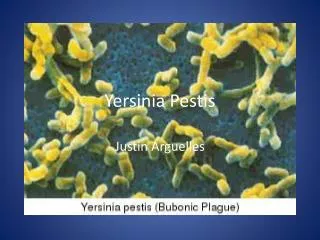
Yersinia. By. Dr. Emad AbdElhameed Morad. Lecturer of Medical Microbiology and Immunology. Yersinia pestis. Morphology. Gram negative, short ovoid, non motile bacilli. In tissues, form capsule like outer envelope.

E N D
Yersinia By Dr. Emad AbdElhameed Morad Lecturer of Medical Microbiology and Immunology
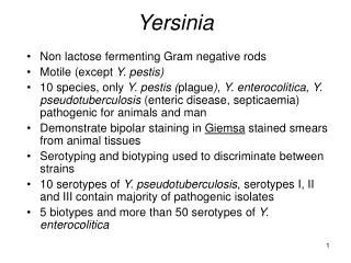
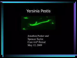
Morphology Gram negative, short ovoid, non motile bacilli. In tissues, form capsule like outer envelope. When stained by Wayson stain or Giemsa, they show marked bipolar staining (Safety pin appearance).
Cultural characters Facultative anaerobes. Grow on MacConkey’s agar producing pale colonies. Grow also on blood agar. The optimum temperature for growth is 27 – 30 degrees. The bacteria is highly pathogenic to white rats. When injected subcutaneously, they die within few days. Films from the liver, spleen and blood show the bacilli.
Biochemical reactions Catalase positive Oxidase negative
Virulence factors F1 antigenis antiphagocytic. V-W antigensare also antiphagocytic. Yersinia outer proteins (Yops)prevent Inflammatory response + antiphagocytic. Plasminogen activator: protease that degrade fibrin facilitating systemic spread of the organism. Lipopolysaccharide endotoxin. Exotoxin which is lethal to mice.
Pathogenesis and clinical picture Yersinia pestis causes plague (black death). Plague is a disease of wild rodents. It is transmitted from rat to rat and from rat to man by the bite of infected rat flea (xenopsylla cheopis).
The inoculated organism multiplies in the draining lymph nodes which become enlarged and tender (bubonic plague). The organism may disseminate to the lung (pneumonic plague).Here, the disease could be transmitted by droplet infection. Dissemination to the blood may occur causing septicaemic plague. Cutaneous hemorrhages may occur so plague is called black death.
Laboratory diagnosis Specimen: aspirate of lymph nodes, sputum, blood. Direct smear stained by Wayson stain to demonstrate bipolarity.
Culture on blood agar & MacConkey’s agar at 27 – 30 degree. Colonies are identified by morphology, biochemical reactions, injection in white rats. Direct detection of F1 antigen in specimen by immunofluorescence. Direct detection of DNA by PCR. Serological diagnosis by ELISA or passive hemagglutination.
Treatment Streptomycin is the drug of choice. Tetracycline or doxycycline is an alternative.
Prophylaxis A formalin killed vaccine was available till 1999, then its production was stopped. New improved vaccines are under development.
It causes enterocolitis. Also, causes mesenteric adenitis which is clinically similar to appendicitis. Transmitted by contamination of food with excreta of domestic animals. Isolation from the stool is done by cold enrichment. Cold enrichment is done by inoculation of the stool in saline incubated at 4 degrees for 2 weeks. Then subculture is done on MacConkey’s agar which will be incubated at 25 degrees.