Download
1 / 60
600 likes | 608 Views
Cells and Cellular Processes Review. Anatomy & Physiology. Objectives. Define all vocabulary terms. (DOK 1) Describe the structure and function of the following cellular structures: cytoplasm, cell membrane, and nucleus. (DOK 1)

E N D
Cells and Cellular Processes Review Anatomy & Physiology
Objectives • Define all vocabulary terms. (DOK 1) • Describe the structure and function of the following cellular structures: cytoplasm, cell membrane, and nucleus. (DOK 1) • Demonstrate why semi-permeable membranes are necessary for biological function, and describe methods of transporting molecules into and out of cells. (DOK 2, 3) • Describe the structure, function and location of the following cellular organelles: ribosomes, cytoskeleton, endoplasmic reticula (rough and smooth), Golgi apparati, peroxisomes, lysosomes, mitochondria. (DOK 1, 2) • Describe the structure and explain the function of various cellular structures for movement, including pseudopodia, flagella, and cilia. (DOK 1, 2) • Explain the structure and function of microvilli, and explain the benefit of microvilli to rate of absorption. (DOK 1, 2, 3) • Compare and contrast active and passive transport, and describe the process of each. (DOK 1, 4) • Explain the structure and function of enzymes, and describe how they operate. (DOK 1, 2) • Analyze the consequences of altering the shape of an enzyme on that enzyme’s function. (DOK 4)
Characteristics of Eukaryotic Cells • First appeared in the fossil record 1.5 billion years ago. • Have a nucleus present. • Have complex membrane bound organelles (mitochondria, chloroplasts, Golgi apparati, etc.) • All members of the following kingdoms have this type of cell: Protista, Fungi, Plantae, Animalia.
Cell Structures, Organelles, and Their Functions Organelles are tiny structures within a cell with specific structures and functions. Three structures that are not considered organelles: • Cytoplasm • Cell membrane • Nucleus
Cell Structures, Organelles, and Their Functions • Cytoplasm: a colloid (thick mixture) material rich in nutrients, enzymes, and water. • All the organelles and internal cellular structures are suspended in this material. • Found in all cell types.
Cell Structures, Organelles, and Their Functions • Cell membrane / Plasmamembrane: • Controls what enters or leaves the cell • Forms the barrier between the cell and its outer environment. • Found in all cell types.
Cell Structures, Organelles, and Their Functions • Nucleus: contains the genetic material (DNA) and controls the cells activities. • Found only in eukaryotic cells. • Composed of: • a porous, double layered outer membrane, the nuclear envelope • Tightly-packed strands of DNA called chromatin • a dark structure involved with the synthesis of RNA called the nucleolus.
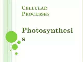
Cell or Plasma Membrane • The function of the cell membrane is to form a barrier between the cell’s inner and outer environment. • It is selectively permeable; only certain materials are able to pass through the membrane. Other materials are blocked.
Cell or Plasma Membrane • It is composed of a phospholipid bilayer with protein molecules embedded within in the bilayer. • Some of these proteins pass completely through both layers of phospholipids. • There are also other types of molecules, such as cholesterol and carbohydrates, that stick off of the outside surface of the cell membrane.
Cell or Plasma Membrane StructureIntegral Proteins and Their Functions
Cytoskeleton • Cytoskeleton: The cytoskeleton is a “framework” that supports the cell membrane and other cell structures within the cytoplasm. • The cytoskeleton is involved with the transport of materials within the cell.
Cytoskeleton Figure 3.24
Microtubules • Dynamic, hollow tubes made of the spherical protein tubulin • Determine the overall shape of the cell and distribution of organelles
Microfilaments • Dynamic strands of the protein actin • Attached to the cytoplasmic side of the plasma membrane • Braces and strengthens the cell surface • Function in endocytosis and exocytosis
Ribosomes • Ribosomes: Ribosomes are the structures within the cell which read m-RNA and assembles amino acids into polypeptide chains. • They are found free floating in the cytoplasm or attached to the nuclear envelope or the rough endoplasmic reticulum.
Endoplasmic Reticulum • Endoplasmic Reticulum (ER): The endoplasmic reticulum is a series of single membrane channels which run throughout the cytoplasm of the cell.
Endoplasmic Reticulum • The smooth endoplasmic reticulum (SER) is free of ribosomes and functions in lipid synthesis, metabolism of carbohydrates, and as a detoxification center of the cell.
Smooth ER • Catalyzes different reactions in different organs of the body • In the liver – lipid and cholesterol metabolism, breakdown of glycogen and, along with the kidneys, detoxification of drugs • In the testes – synthesis of steroid-based hormones • In the intestinal cells – absorption, synthesis, and transport of fats • In skeletal and cardiac muscle – storage and release of calcium
Endoplasmic Reticulum • The rough endoplasmic reticulum (RER) has ribosomes attached to its outer membrane layer and is the site of protein synthesis. • These are secretory proteins, which will be secreted (released) by the cell.
Ribosome mRNA 1 As the protein is synthesized on the ribosome, it migrates into the rough ER cistern. Rough ER 1 Protein Figure 3.5, step 1
Ribosome mRNA 1 As the protein is synthesized on the ribosome, it migrates into the rough ER cistern. Rough ER In the cistern, the protein folds into its functional shape. Short sugar chains may be attached to the protein (forming a glycoprotein). 2 1 2 Protein Figure 3.5, step 2
Ribosome mRNA 1 As the protein is synthesized on the ribosome, it migrates into the rough ER cistern. Rough ER In the cistern, the protein folds into its functional shape. Short sugar chains may be attached to the protein (forming a glycoprotein). 2 1 3 2 Protein The protein is packaged in a tiny membranous sac called a transport vesicle. 3 Transport vesicle buds off Figure 3.5, step 3
Ribosome mRNA 1 As the protein is synthesized on the ribosome, it migrates into the rough ER cistern. Rough ER In the cistern, the protein folds into its functional shape. Short sugar chains may be attached to the protein (forming a glycoprotein). 2 1 3 2 Protein The protein is packaged in a tiny membranous sac called a transport vesicle. 3 4 Transport vesicle buds off The transport vesicle buds from the rough ER and travels to the Golgi apparatus for further processing. 4 Protein inside transport vesicle Figure 3.5, step 4
Golgi Apparatus • Golgi Apparatus: The Golgi apparatus looks like a series of flattened, stacked, membrane sacs. • The Golgi apparatus is the center for manufacturing, modifying, and packaging of materials for transport.
Golgi Apparatus • Proteins to be packaged are received from the RER on the cis (flattened) face • There the proteins are tagged and packaged in small bubbles of membrane called secretory vesicles • The vesicles pinch off the trans(rounded, with bubbles) face of the organelle structure. • Cells, such as those found in gland tissues, contain MANY Golgi apparati.
Golgi Apparatus Rough ER Cisterna Proteins in cisterna Lysosome fuses with ingested substances Membrane Transport vesicle Golgi vesicle containing digestive enzymes becomes a lysosome Pathway 3 Pathway 2 Golgi apparatus Golgi vesicle containing membrane components fuses with the plasma membrane Secretory vesicles Pathway 1 Proteins Golgi vesicle containing proteins to be secreted becomes a secretory vesicle Plasma membrane Secretion by exocytosis Extracellular fluid Figure 3.6
Lysosomes Lysosomes: membrane bound sacs containing strong enzymes produced by ribosomes • digest all forms of macromolecules. (large molecules) • Packaged by the Golgi apparatus • recycle old cell organelles and structures. • If the cell is damaged or infected by a pathogen, lysosomes will rupture, and destroy the cell in the process. • Thus giving them the name “suicide bags”.
Lysosomes in the Body • Digest ingested bacteria, viruses, and toxins • Breakdown glycogen and release thyroid hormone • Breakdown nonuseful tissue • Breakdown bone to release Ca2+ • Secretory lysosomes are found in white blood cells, immune cells, and melanocytes
Peroxisomes • Peroxisomes: special vesicles (bubbles) made from a single layer of membrane. • Involved with the metabolism of lipids and the detoxification of certain substances such as alcohol.
Peroxisomes • Produce H2O2 (peroxide) during the detox reaction, which gives them their name. • H2O2 is also toxic to the cell, but peroxisomes contain an enzyme called catalase which breaks down the H2O2 into H2O and O2 molecules which are nontoxic.
Mitochondria • Mitochondria: a double membrane structure. The outer membrane surrounds a highly folded inner membrane. The folds are called cristae. • The site of aerobic cellular respiration, by which ATP is produced.
Mitochondria • The inner space within the mitochondrion is called the matrix, and contains cytoplasm, ribosomes, and DNA. • Mitochondria are sometimes called “powerhouse of the cell”
Cellular Projections (Not found in all cells) Flagella propel the cell The only flagellated cell in the human body is sperm Cilia move materials across the cell surface Located in the respiratory system to move mucus Pseudopodiaprojections of the cell membrane used for movement and digestion Microvilli are tiny, fingerlike extensions of the plasma membrane Increase surface area for absorption
Cellular Movement 1. Flagella 2. Cillia 3. Pseudopod
2. Cilia • Whiplike, motile cellular extensions on exposed surfaces of certain cells • Move substances in one direction across cell surfaces
2. Cilia Figure 3.27c
3. Pseudopodia • Pseudopodia literally means false foot. • Extensions of the cell’s cytoplasm • Used for movement • Also used for phagocytosis (“cell eating”, from Ancient Greek), a process for taking in molecules that are too large to pass through the cell membrane. • White blood cells which engulf (completely surround) and destroy bacteria and dead cell materials are called phagocytes.
4. Microvilli • Microvilli are tiny finger-like projections on certain types of cells. • They function to increase the rate of absorption of materials, because they increase the surface area of the cell membrane. • They are found on the surface of the cells that line the small intestine of animals.
4. Microvilli microvilli on apical (top) surface of intestinal cells
Developmental Aspects of Cells • All cells of the body contain the same DNA BUT develop into different specialized cells • Cells in various parts of the embryo are exposed to different chemical signals that cause them to develop differently • Genes of specific cells are turned on or off (i.e., by methylation of their DNA)
Developmental Aspects of Cells • Turning genes on and off changes which proteins are made by the cell (among other things). • Cell specialization is determined by the kind of proteins that are made in that cell • Development of specific and distinctive features in cells is called cell differentiation
Developmental Aspects of Cells • Cell aging • Wear and tear theory attributes aging to the accumulation of chemical damage and formation of free radicals that have cumulative effects throughout life • Genetic theory attributes aging to a cessation (stopping) of mitosis that is programmed into our genes
Example Cell Types – Connective tissue Fibroblasts Rough ER and Golgi apparatus No organelles Nucleus Erythrocytes (a) Cells that connect body parts
Example Cell Types – Epithelial Cells (line cavities and cover organs) Nucleus Epithelial cells Intermediate filaments (b) Cells that cover and line body organs Figure 3.8b
Example Cell Types – Muscle Cells Skeletal muscle cell Nuclei Contractile filaments Smooth muscle cells (c) Cells that move organs and body parts Figure 3.8c
Lipid droplet Fat cell Example Cell Types – Adipose cells (Fat storage cells) Nucleus (d) Cell that stores nutrients Figure 3.8d