Download
1 / 4
40 likes | 47 Views
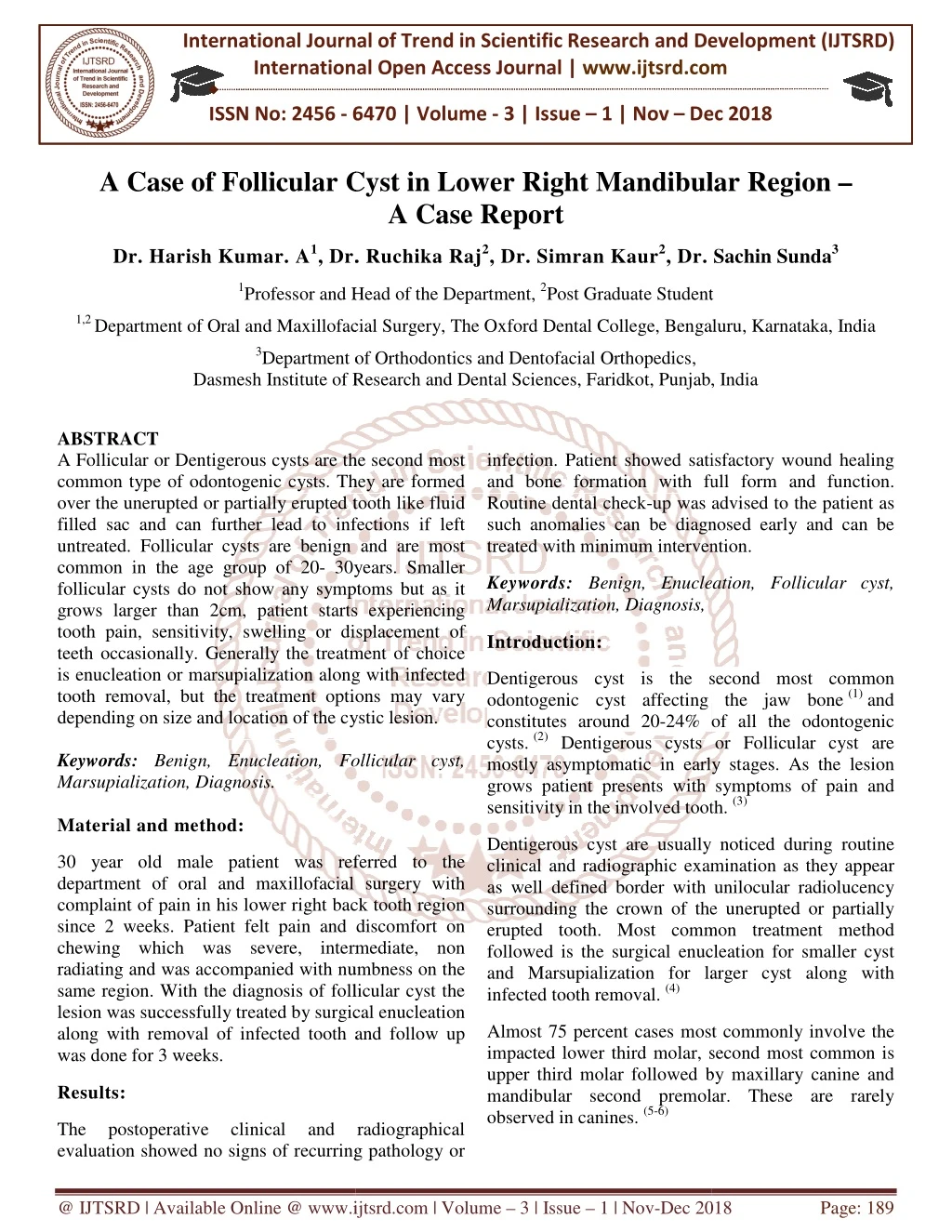
A Follicular or Dentigerous cysts are the second most common type of odontogenic cysts. They are formed over the unerupted or partially erupted tooth like fluid filled sac and can further lead to infections if left untreated. Follicular cysts are benign and are most common in the age group of 20 30years. Smaller follicular cysts do not show any symptoms but as it grows larger than 2cm, patient starts experiencing tooth pain, sensitivity, swelling or displacement of teeth occasionally. Generally the treatment of choice is enucleation or marsupialization along with infected tooth removal, but the treatment options may vary depending on size and location of the cystic lesion. Dr. Harish Kumar. A | Dr. Ruchika Raj | Dr. Simran Kaur | Dr. Sachin Sunda "A Case of Follicular Cyst in Lower Right Mandibular Region A Case Report" Published in International Journal of Trend in Scientific Research and Development (ijtsrd), ISSN: 2456-6470, Volume-3 | Issue-1 , December 2018, URL: https://www.ijtsrd.com/papers/ijtsrd18958.pdf Paper URL: http://www.ijtsrd.com/medicine/other/18958/a-case-of-follicular-cyst-in-lower-right-mandibular-region-u00e2u20acu201c-a-case-report/dr-harish-kumar-a<br>

E N D
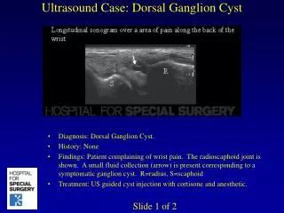
International Journal of Trend in International Open Access Journal International Open Access Journal | www.ijtsrd.com International Journal of Trend in Scientific Research and Development (IJTSRD) Research and Development (IJTSRD) www.ijtsrd.com ISSN No: 2456 - 6470 6470 | Volume - 3 | Issue – 1 | Nov – Dec 2018 Dec 2018 A Case of Follicular Cyst in A Case of Follicular Cyst in Lower Right Mandibular Region A Case Report Lower Right Mandibular Region – Dr. Harish Kumar. A1, Dr. , Dr. Ruchika Raj2, Dr. Simran Kaur2, Dr. Sachin Sunda3 1Professor and Head of the Department, Professor and Head of the Department, 2Post Graduate Student tudent 1,2 Department of Oral and Maxillofacial axillofacial Surgery, The Oxford Dental College, Bengaluru Bengaluru, Karnataka, India 3Department of Orthodontics and Dentofacial Orthopedics, Dasmesh Institute of Research and Dental Sciences, Dasmesh Institute of Research and Dental Sciences, Faridkot, Punjab, India Department of Orthodontics and Dentofacial Orthopedics, , India ABSTRACT A Follicular or Dentigerous cysts are the second most common type of odontogenic cysts. They are formed over the unerupted or partially erupted tooth like fluid filled sac and can further lead to infections if left untreated. Follicular cysts are benign and are most common in the age group of 20- 30years. Smaller follicular cysts do not show any symptoms but as it grows larger than 2cm, patient starts experiencing tooth pain, sensitivity, swelling or displacement of teeth occasionally. Generally the treatment of choice is enucleation or marsupialization along with infected tooth removal, but the treatment options may vary depending on size and location of the cystic lesion. Keywords: Benign, Enucleation, Follicular cyst, Marsupialization, Diagnosis. Material and method: infection. Patient showed satisfactory wound healing and bone formation with full form and function. Routine dental check-up was advised to the patient as such anomalies can be diagnosed early and can be treated with minimum intervention. treated with minimum intervention. A Follicular or Dentigerous cysts are the second most common type of odontogenic cysts. They are formed or partially erupted tooth like fluid filled sac and can further lead to infections if left untreated. Follicular cysts are benign and are most infection. Patient showed satisfactory wound healing and bone formation with full form and function. up was advised to the patient as diagnosed early and can be 30years. Smaller Keywords: Benign, Enucleation, Follicular cyst, Marsupialization, Diagnosis, Benign, Enucleation, Follicular cyst, follicular cysts do not show any symptoms but as it r than 2cm, patient starts experiencing tooth pain, sensitivity, swelling or displacement of teeth occasionally. Generally the treatment of choice is enucleation or marsupialization along with infected tooth removal, but the treatment options may vary nding on size and location of the cystic lesion. Introduction: Dentigerous cyst is the second most common odontogenic cyst affecting the jaw bone (1) and 24% of all the odontogenic Dentigerous cysts or Follicular cyst are mostly asymptomatic in early stages. As the lesion grows patient presents with symptoms of pain and Dentigerous cyst is the second most common odontogenic cyst affecting the jaw bone constitutes around 20-24% of all the odontogenic cysts. (2) Dentigerous cysts or Follicular cyst are mostly asymptomatic in early stages. As the lesion grows patient presents with symptoms of pain and sensitivity in the involved tooth. sensitivity in the involved tooth. (3) Benign, Enucleation, Follicular cyst, Dentigerous cyst are usually clinical and radiographic examination as they appear as well defined border with unilocular radiolucency surrounding the crown of the unerupted or partially erupted tooth. Most common treatment method followed is the surgical enucle and Marsupialization for larger cyst along with infected tooth removal. (4) noticed during routine 30 year old male patient was referred to the department of oral and maxillofacial surgery with complaint of pain in his lower right back tooth region since 2 weeks. Patient felt pain and discomfort on chewing which was severe, intermediate, non radiating and was accompanied with numbness on the same region. With the diagnosis of follicular cyst the lesion was successfully treated by surgical enucleation along with removal of infected tooth and follow up was done for 3 weeks. 30 year old male patient was referred to the department of oral and maxillofacial surgery with complaint of pain in his lower right back tooth region since 2 weeks. Patient felt pain and discomfort on chewing which was severe, intermediate, non nd was accompanied with numbness on the same region. With the diagnosis of follicular cyst the lesion was successfully treated by surgical enucleation along with removal of infected tooth and follow up clinical and radiographic examination as they appear as well defined border with unilocular radiolucency surrounding the crown of the unerupted or partially erupted tooth. Most common treatment method followed is the surgical enucleation for smaller cyst and Marsupialization for larger cyst along with Almost 75 percent cases most commonly involve the impacted lower third molar, second most common is upper third molar followed by maxillary canine and bular second premolar. These are rarely Almost 75 percent cases most commonly involve the impacted lower third molar, second most common is upper third molar followed by maxillary canine and mandibular second premolar. These are rarely observed in canines. (5-6) Results: The evaluation showed no signs of recurring pathology or evaluation showed no signs of recurring pathology or postoperative clinical cal and and radiographical radiographical @ IJTSRD | Available Online @ www.ijtsrd.com www.ijtsrd.com | Volume – 3 | Issue – 1 | Nov-Dec 2018 Dec 2018 Page: 189
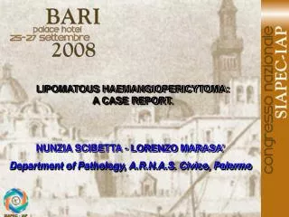
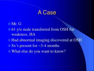
International Journal of Trend in Scientific Research and Development (IJTSRD) International Journal of Trend in Scientific Research and Development (IJTSRD) International Journal of Trend in Scientific Research and Development (IJTSRD) ISSN: 2456-6470 Case report: Provisional Diagnosis: A 30 year old male patient visited the department of oral and maxillofacial surgery, the Oxford Dental College for evaluation of the pain and discomfort in his lower right back tooth region. Patient gave history of pain as insidious in onset, intermittent in nature, severe in intensity, aggravates on chewing and gets relives by its own. A 30 year old male patient visited the department of oral and maxillofacial surgery, the Oxford Dental College for evaluation of the pain and discomfort in back tooth region. Patient gave history of pain as insidious in onset, intermittent in nature, severe in intensity, aggravates on chewing and gets Radicular cyst, eruption cyst and dentigerous cyst. Radicular cyst, eruption cyst and dentigerous cyst. Treatment plan: Based on the history and correlating it with the radiographic and clinical evaluation, a conservative treatment plan of surgical enucleation of the cystic lesion with 2mm of healthy bone and tissue was performed along with removal of involved tooth (46, 47, and 48) under general anaesthesia. The specimen was sent for histological evaluation. Irrigation of infected area was done with saline and the bony edges were smoothened. Primary haemostasis was achieved and wound was sutured with further dressing. No bone grafts were used for the surgical bed as the idea was to initiate primary wound healing where the clot wou for osseous remodelling and growth. Patient was kept on antibiotics and analgesics for 5 days. The excised mass comprised of cyst along with cystic lining of tissues and the involved tooth (figure 2) tissues and the involved tooth (figure 2) Based on the history and correlating it with the radiographic and clinical evaluation, a conservative enucleation of the cystic lesion with 2mm of healthy bone and tissue was performed along with removal of involved tooth (46, 47, and 48) under general anaesthesia. The specimen was sent for histological evaluation. Irrigation of infected area was done with betadine and normal saline and the bony edges were smoothened. Primary haemostasis was achieved and wound was sutured with further dressing. No bone grafts were used for the surgical bed as the idea was to initiate primary wound healing where the clot would act as a scaffold for osseous remodelling and growth. Patient was kept on antibiotics and analgesics for 5 days. The excised mass comprised of cyst along with cystic lining of xtra-oral examination: Gross asymmetry of the face was observed on lower right side of face. The overlying skin showed no signs of inflammation. The swelling measured 4*3cm approximately (length* width) and was tender on palpation with bony hard consistency. Lymph nodes were non- palpable and non-tender. No Neurological deficit was noted on right side of face. Gross asymmetry of the face was observed on lower ace. The overlying skin showed no signs of inflammation. The swelling measured 4*3cm approximately (length* width) and was tender on palpation with bony hard consistency. Lymph nodes tender. No Neurological Intra-oral examination: Revealed a hard- tender swelling in the lower right vestibular region extending from distal of lower right first molar to 3-4cm distal to second molar extending to ramus region measuring 5 cm*3cm*3cm in length, width and height respectively. Swelling was tender on palpation and erythematous with occasional pus discharge from the pocket distal to second molar. Teeth (47, 46) were tender on percussion, pulp vitality was performed to check the vitality of 46 and 47 t rule out any underlying periapical/ periodontal pathology and the teeth were vital. Vestibular obliteration was observed from distal to first molar. On palpation there was expansion of buccal and cortical plates extending distal to 46. tender swelling in the lower right vestibular region extending from distal of lower right 4cm distal to second molar extending to ramus region measuring 5 cm*3cm*3cm in length, idth and height respectively. Swelling was tender on palpation and erythematous with occasional pus discharge from the pocket distal to second molar. Teeth (47, 46) were tender on percussion, pulp vitality was performed to check the vitality of 46 and 47 to rule out any underlying periapical/ periodontal pathology and the teeth were vital. Vestibular obliteration was observed from distal to first molar. On palpation there was expansion of buccal and Histological evaluation: (figure 3 Histological evaluation: (figure 3) •The epithelium was 3 was non -keratinized with irregularly arranged flat end cells. •No retepeg formation was noticed. •Primarily the specimen fibroblastic tissue mucopolysaccharide entrapments. •Linear and curved hematogenous bodies were noticed which resembles rushton bodies. •Inflammatory infiltrate neutrophils were present in the watery blood tinged aspirate. •Incompletely formed 48 (incomplete root formation) in the cystic cavity circumferential variety of dentigerous cyst. circumferential variety of dentigerous cyst. The epithelium was 3-4 layers thick which keratinized with irregularly arranged No retepeg formation was noticed. Primarily the fibroblastic mucopolysaccharide entrapments. specimen tissue consisted which which consisted of of had had hematogenous bodies were noticed which resembles rushton bodies. Inflammatory infiltrate – lymphocytes and neutrophils were present in the watery blood Differential diagnosis: Periapical cyst, eruption cyst, odontogenic keratocyst, radicular cyst Periapical cyst, eruption cyst, odontogenic keratocyst, Incompletely formed 48 (incomplete root formation) in the cystic cavity- resembling a Radiographic evaluation: (fig 1) Follow-up: OPG revealed unilocular radiolucent lesion with thin well-defined radio-opaque schlerotic border extending posteriorly 2 cm away from the roots of impacted tooth (48), anteriorly involving the mesio buccal root of first molar (46), inferiorly 1.5 cm above the lower border of mandible and superiorly 1.8 cm below the alveolar ridge. Obliteration of mandibular canal was noted with loss of normal trabecular pattern of bone. OPG revealed unilocular radiolucent lesion with thin opaque schlerotic border extending posteriorly 2 cm away from the roots of inverted impacted tooth (48), anteriorly involving the mesio- buccal root of first molar (46), inferiorly 1.5 cm above the lower border of mandible and superiorly 1.8 cm below the alveolar ridge. Obliteration of mandibular Follow up was done for 3 weeks. Post operatively OPG did not show any signs of recurrence or infection. satisfactory wound healing was observed Follow up was done for 3 weeks. Post operatively OPG did not show any signs of recurrence or infection. satisfactory wound healing was observed clinically. Discussion: al trabecular pattern Dentigerous or follicular cyst is the most common type of non -inflammatory odontogenic cyst. Dentigerous cyst is an entity derived from remnants of Dentigerous cyst is an entity derived from remnants of or follicular cyst is the most common inflammatory odontogenic cyst. @ IJTSRD | Available Online @ www.ijtsrd.com www.ijtsrd.com | Volume – 3 | Issue – 1 | Nov-Dec 2018 Dec 2018 Page: 190
International Journal of Trend in Scientific Research and Development (IJTSRD) International Journal of Trend in Scientific Research and Development (IJTSRD) International Journal of Trend in Scientific Research and Development (IJTSRD) ISSN: 2456-6470 dental organ and reduced enamel epithelium and is attached to crown of the unerupted or partially erupted tooth (1) dental organ and reduced enamel epithelium and is attached to crown of the unerupted or partially erupted dentigerous cyst associated with an impacted mandibular canine. Dentistry and Medical Research. 2014 Jul 1;2(2):49. 6)Bhange P, Sayed Z, Irani M. A Typical Dentigerous Cyst in the Mandible. Journal of Medical and Dental 2016;3(6):40-3. 7)Tümer C, Eset AE, Atabek A. Ectopic impacted mandibular third molar in the subcondylar region associated with a dentigerous cyst: A case report. Quintessence international. 2002 Mar 1;33(3). 8)Mohapatra PK, Joshi management of a dentigerous cyst associated with an impacted mandibular second premolar in mixed. Journal of dental research, dental clinics, dental prospects. 2009;3(3):98. dental prospects. 2009;3(3):98. dentigerous cyst associated with an impacted mandibular canine. Dentistry and Medical Research. 2014 Jul 1;2(2):49. Bhange P, Sayed Z, Irani M. A Typical Dentigerous Cyst in the Mandible. Journal of Medical and Dental Different variants of dentigerous or follicular cyst has been reported like central type, lateral type and circumferencial type which involves the whole tooth and are difficult to differentiate from keratocyst odontogenic tumour hence, examination is mandatory for confirmation of the diagnosis(6) fferent variants of dentigerous or follicular cyst has been reported like central type, lateral type and circumferencial type which involves the whole tooth and are difficult to differentiate from keratocyst odontogenic tumour hence, ination is mandatory for confirmation of the Science Science Research. Research. histopathological histopathological Tümer C, Eset AE, Atabek A. Ectopic impacted mandibular third molar in the subcondylar region associated with a dentigerous cyst: A case report. Quintessence international. 2002 Mar 1;33(3). Treatment modalities like marsupialization with iodoform or enucleation are preferred depending on the size of the lesion. (7) Main aim of treating dentigerous cyst is complete removal of the pathol with minimal surgical intervention. Treatment modalities like marsupialization with iodoform or enucleation are preferred depending on the size of the lesion. (7) Main aim of treating dentigerous cyst is complete removal of the pathology , Joshi N. N. Conservative Conservative management of a dentigerous cyst associated with an impacted mandibular second premolar in mixed. Journal of dental research, dental clinics, Conclusion: The presented case is of follicular or dentigerous cyst in a 30 year old male patient with inverted and impacted third molar 48. These types of cases should not be diagnosed only on basis of clinical moda Radiographic and histopathological examination is mandatory for confirmation of the underlying pathology and follow up of the patient should be carried out to check for any recurrence and infection. Conservative treatment methods with minimal intervention should always be the priority for smaller cystic lesion. The presented case is of follicular or dentigerous cyst in a 30 year old male patient with inverted and impacted third molar 48. These types of cases should not be diagnosed only on basis of clinical modality. Radiographic and histopathological examination is mandatory for confirmation of the underlying pathology and follow up of the patient should be carried out to check for any recurrence and infection. Conservative treatment methods with minimal ntion should always be the priority for smaller FIGURE 1: Pre Operative OPG Of Patient Depicting FIGURE 1: Pre Operative OPG Of Radiolucent Lesion Involving 46, 47 and 48. Radiolucent Lesion Involving 46, 47 and 48. References: 1)Rajendran R. Shafer's textbook of oral pathology. Elsevier India; 2009. 2)Daley TD, Wysocki GP, Pringle GA. Relative incidence of odontogenic tumors and oral and jaw cysts in a Canadian population. Oral Surgery, Oral Medicine, Oral Pathology. 1994 Mar 1;77(3):276 80. 3)Goel A, Patil P, Bansal R, Sabharwal R. Dentigerous cyst involving mandibular third molar: conservative treatment with radiologic follow-up and review of literature. Clini Investigation Journal. 2013 Jul 1;2(3):233. 4)Dunfee BL, Sakai O, Pistey R, Gohel A. Radiologic and pathologic characteristics of benign and malignant lesions of the mandible. Radiographics. 2006 Nov;26(6):1751 5)Yaqoob A, Wani TM, Ashraf J, Yaqoob G, Yaqub N. Conservative surgical management of a N. Conservative surgical management of a Rajendran R. Shafer's textbook of oral pathology. Daley TD, Wysocki GP, Pringle GA. Relative incidence of odontogenic tumors and oral and jaw an population. Oral Surgery, Oral Medicine, Oral Pathology. 1994 Mar 1;77(3):276- FIGURE 2: Clinical Picture of The Excised Lesion FIGURE 2: Clinical Picture of The Excised Lesion Goel A, Patil P, Bansal R, Sabharwal R. Dentigerous cyst involving mandibular third molar: conservative treatment with radiologic up and review of literature. Clinical Cancer Investigation Journal. 2013 Jul 1;2(3):233. Dunfee BL, Sakai O, Pistey R, Gohel A. Radiologic and pathologic characteristics of benign and malignant lesions of the mandible. Radiographics. 2006 Nov;26(6):1751-68. Yaqoob G, Yaqub @ IJTSRD | Available Online @ www.ijtsrd.com www.ijtsrd.com | Volume – 3 | Issue – 1 | Nov-Dec 2018 Dec 2018 Page: 191
International Journal of Trend in Scientific Research and Development (IJTSRD) International Journal of Trend in Scientific Research and Development (IJTSRD) International Journal of Trend in Scientific Research and Development (IJTSRD) ISSN: 2456-6470 FIGURE 3: Histopatholical picture of follicular cyst FIGURE 3: Histopatholical picture of follicular cyst @ IJTSRD | Available Online @ www.ijtsrd.com www.ijtsrd.com | Volume – 3 | Issue – 1 | Nov-Dec 2018 Dec 2018 Page: 192