Download
1 / 16
660 likes | 3.6k Views
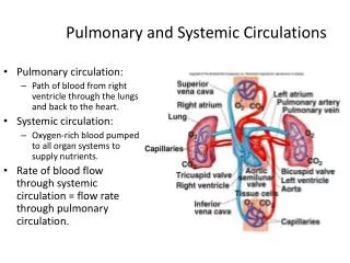
Pulmonary and Systemic Circulations. Pulmonary circulation: Path of blood from right ventricle through the lungs and back to the heart. Systemic circulation: Oxygen-rich blood pumped to all organ systems to supply nutrients.

E N D
Pulmonary and Systemic Circulations • Pulmonary circulation: • Path of blood from right ventricle through the lungs and back to the heart. • Systemic circulation: • Oxygen-rich blood pumped to all organ systems to supply nutrients. • Rate of blood flow through systemic circulation = flow rate through pulmonary circulation.
Role is to direct the flow of blood from the heart to the capillaries, and back to the heart. Arteries. Arterioles. Capillaries. Venules. Veins. Systemic Circulation
Blood Vessels • Walls composed of 3 “tunics:” • Tunica externa: • Outer layer comprised of connective tissue. • Tunica media: • Middle layer composed of smooth muscle. • Tunica interna: • Innermost simple squamous endothelium. • Basement membrane. • Layer of elastin.
Blood Vessels (continued) • Elastic arteries: • Numerous layers of elastin fibers between smooth muscle. • Expand when the pressure of the blood rises. • Act as recoil system when ventricles relax. • Muscular arteries: • Are less elastic and have a thicker layer of smooth muscle. • Diameter changes slightly as BP raises and falls. • Arterioles: • Contain highest % smooth muscle. • Greatest pressure drop. • Greatest resistance to flow.
Blood Vessels (continued) • Most of the blood volume is contained in the venous system. • Venules: • Formed when capillaries unite. • Very porous. • Veins: • Contain little smooth muscle or elastin. • Capacitance vessels (blood reservoirs). • Contain 1-way valves that ensure blood flow to the heart. • Skeletal muscle pump and contraction of diaphragm: • Aid in venous blood return of blood to the heart.
Types of Capillaries • Capillaries: • Smallest blood vessels. • 1 endothelial cell thick. • Provide direct access to cells. • Permits exchange of nutrients and wastes. • Continuous: • Adjacent endothelial cells tightly joined together. • Intercellular channels that permit passage of molecules (other than proteins) between capillary blood and tissue fluid. • Muscle, lungs, and adipose tissue. • Fenestrated: • Wide intercellular pores. • Provides greater permeability. • Kidneys, endocrine glands, and intestines. • Discontinuous (sinusoidal): • Have large, leaky capillaries. • Liver, spleen, and bone marrow.
Atherosclerosis • Most common form of arteriosclerosis (hardening of the arteries). • Mechanism of plaque production: • Begins as a result of damage to endothelial cell wall. • HTN, smoking, high cholesterol, and diabetes. • Cytokines are secreted by endothelium; platelets, macrophages, and lymphocytes. • Attract more monocytes and lymphocytes.
Atherosclerosis (continued) • Monocytes become macrophages. • Engulf lipids and transform into foam cells. • Smooth muscle cells synthesize connective tissue proteins. • Smooth muscle cells migrate to tunica interna, and proliferate forming fibrous plaques.
Cholesterol and Plasma Lipoproteins • High blood cholesterol associated with risk of atherosclerosis. • Lipids are carried in the blood attached to protein carriers. • Cholesterol is carried to the arteries by LDLs (low-density lipoproteins). • LDLs are produced in the liver. • LDLs are small protein-coated droplets of cholesterol, neutral fat, free fatty acids, and phospholipids.
Cholesterol and Plasma Lipoproteins (continued) • Cells in various organs contain receptors for proteins in LDL. • LDL protein attaches to receptors. • The cell engulfs the LDL and utilizes cholesterol for different purposes. • LDL is oxidized and contributes to: • Endothelial cell injury. • Migration of monocytes and lymphocytes to tunica interna. • Conversion of monocytes to macrophages. • Excessive cholesterol is released from the cells. • Travel in the blood as HDLs (high-density lipoproteins), and removed by the liver. • Artery walls do not have receptors for HDL.
Ischemic Heart Disease • Ischemia: • Oxygen supply to tissue is deficient. • Most common cause is atherosclerosis of coronary arteries. • Increased [lactic acid] produced by anaerobic respiration. • Angina pectoris: • Substernal pain. • Myocardial infarction (MI): • Changes in T segment of ECG. • Increased CPK and LDH.
Arrhythmias Detected on ECG • Arrhythmias: • Abnormal heart rhythms. • Flutter: • Extremely rapid rates of excitation and contraction of atria or ventricles. • Atrial flutter degenerates into atrial fibrillation. • Fibrillation: • Contractions of different groups of myocardial cells at different times. • Coordination of pumping impossible. • Ventricular fibrillation is life-threatening.
Arrhythmias Detected on ECG (continued) • Bradycardia: • HR slower < 60 beats/min. • Tachycardia: • HR > 100 beats/min. • First–degree AV nodal block: • Rate of impulse conduction through AV node exceeds 0.2 sec. • P-R interval. • Second-degree AV nodal block: • AV node is damaged so that only 1 out of 2-4 atrial APs can pass to the ventricles. • P wave without QRS.
Arrhythmias Detected on ECG (continued) • Third-degree (complete) AV nodal block: • None of the atrial waves can pass through the AV node. • Ventricles paced by ectopic pacemaker.
Lymphatic System • 3 basic functions: • Transports interstitial (tissue) fluid back to the blood. • Transports absorbed fat from small intestine to the blood. • Helps provide immunological defenses against pathogens.
Lymphatic System (continued) • Lymphatic capillaries: • Closed-end tubules that form vast networks in intercellular spaces. • Lymph: • Fluid that enters the lymphatic capillaries. • Lymph carried from lymph capillaries, to lymph ducts, and then to lymph nodes. • Lymph nodes filter the lymph before returning it to the veins.