Download
1 / 48
850 likes | 2.21k Views
Ebstein’s anomaly. History. 1866 – Dr. Wilhelm Ebstein described cardiac findings of 19 y.o. patient who had died of cyanotic heart disease 1950 – Helen Taussig - first clinical syndrome analysis

E N D
History 1866 – Dr. Wilhelm Ebstein described cardiac findings of 19 y.o. patient who had died of cyanotic heart disease 1950 – Helen Taussig - first clinical syndrome analysis 1950’s – BT shunt for neonatal Ebstein ( functional tricuspid or pulmonary atresia) 1954 – Wright, Kirklin – direct closure of ASD for correction of right-to-left shunt (patient survived)
1958 – tricuspid valve reconstruction – Hunter & Lillehei – attempt to create competent valve by repositioning of displaced leaflet & excluding atrialized chamber ( 2 patients – both didn’t survive due to CAVB) 1964 - Hardy revived Hunter-Lillehei operation – effective only for mild anomaly; complications: CAVB; RCA injury; RV aneurysm Barnard (1963); Lillehei (1967) – tricuspid valve replacement Danielson (1972); Carpenter (1988) – TV repair based on use of anterior leaflet Starnes (1991) – single ventricle palliation of neonatal Ebstein Knott-Craig (1994) – biventricular repair of neonatal Ebstein anomaly
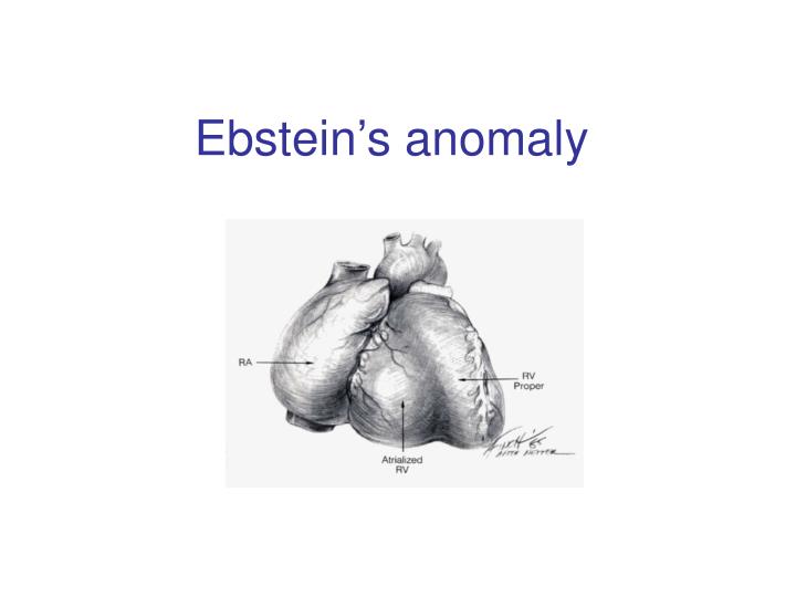
Anatomy Distal attachment of the septal & posterior leaflets away from the atrio-ventricular junction Plane of closure of the tricuspid valve at the junction of the inlet and apical component of the right ventricle Dilatation of the atrio-ventricular junction
Valve pathology is variable from patient to patient Variability of location of the valve annular attachment (from inlet to outlet) Variability of degree of formation and delamination of the septal and posterior leaflet In most cases TV has bifoliate structure with combined antero-superior and posterior leaflet
TV anatomy TV leaflet malformation: Septal > Mural (posterior) >Anterior TV displacement: maximal – postero-septal comissure minimal – antero-septal comissure
Anterior leaflet Enlarged Sail-like Thicken and partially muscularized
Anterior leaflet Leading edge: - free and mobile - segmental direct attachment to the myocardium - linear direct attachment (entire leading edge attached to the myocardium) Delamination: - partial - complete
Antero-superior leaflet of the TV Attachment of the antero-superior leaflet – focal attachment to the medial and anterior papillary muscle
Antero-superior leaflet of the TV Attachment of the antero-superior leaflet – entire leading edge of the leaflet is attached linearly to a muscle between inlet and apical component of RV
Antero-superior leaflet of the TV Edge of the anterior leaflet is attached in hyphenated fashion
Right ventricle Atrialized RV – inlet part of RV above TV attachment; in symptomatic patient tend to be thin-walled and dilated Functional RV – apical and infundibular component: - thinner - contain fewer than normal muscular fibers - contain more fibrous tissue
Carpentier classification (1988) Grade A: mobile anterior leaflet/small contractile atrialized right ventricle Grade B: mobile anterior leaflet/large, noncontractile atrialized RV Grade C: tethering of anterior leaflet/large, noncontractile atrialized RV Grade D: leaflets forming a continuous sac adherent to the right ventricle
Great Ormond Street scorecombined area of the right atrium and atrialized portion of the right ventricle divided by the area of functional RV added to the area of the left heart chambers ( in diastole) Grade 1 – ratio less than 0.5 Grade 2 – ratio 0.5 – 0.99 Grade 3 – ratio 1 – 1.49 Grade 4 – greater than 1.5
Physiology effective pulmonary blood flow Decrease flow through the right heart shunt direction through ASD/PFO Tricuspid valve: - regurgitation - stenosis - stenosis+regurgitation Right ventricle: - functional dysfunction - Anatomical dysfunction
Ebstein’s anomaly Age of presentation depends on severity of tricuspid and RV dysfunction Neonatal Adult
Newborn presentation Functional (anatomical?) pulmonic atresia Congestive heart failure + RVOTO Congestive heart failure PDA dependence TV dysfunction RV dysfunction Elevated PVR
Treatment protocol for Ebstein’s anomaly in the neonate PG E dependent Successful wean Failure to wean No treatment cyanosis Cyanosis + CHF CHF Shunt+TV procedure TV procedure Shunt alone Single ventricle (RV exclusion) TV repair/replacement 1 ½ (BDG+TVR) RV exclusion TV repair/replacement How I Manage Neonatal Ebstein’s Anomaly Edward L. Bove, Jennifer C. Hirsch, Richard G. Ohye, and Eric J. Devaney
Univentricular approach (Starnes procedure, 1991)Indication: TV not amenable to repair Functional portion of TV is inadequate RVOT obstruction
Starnes procedure TV closure with fenestrated ( 4 mm) patch at the anatomic level of the tricuspid annulus. Atrial septectomy RVOT procedure – only PA division for patient with pulmonary artery insufficiency. Reduction atrioplasty BT shunt
Biventricular repair of neonates and infants Treatment concept – biventricular repair versus single-ventricle palliation
Biventricular repair (Knott-Craig, 1994)surgical technique: Reduction right atrioplasty ( true atrium) TV repair: - reduction annuloplasty (annulus 12-14 mm) - construction of monoleaflet TV - augmentation of the functional leaflet if deficient ASD closure with 3 - 5 mm fenestration: unload RV; increase cardiac output. Size of the fenestration inversely proportional to the effectiveness of TV repair. Creation of functional RVOT – use a small patch( RVOT – 7-8 mm in neonate);pulmonary insufficiency is very poorly tolerated Patients with suboptimal TV repair – RVOT should be repaired with valve conduit
Our institutional bias has been to do a two-ventricle repair in all such patients. Between 1994 and 2006, 27 consecutive patients with Ebstein’s anomaly underwent operation.
Table 1. Associated Comorbidities Comorbidity n Pulmonary atresia (functional 7, anatomic 11) 18 Ventricular septal defect 3 Small left ventricle 3 Hypoplastic branch pulmonary arteries 3 Previous cardiac surgery 4 Grade III or IV intracranial hemorrhage 3 Ischemic hepatic necrosis and renal failure creatinine 1.5) 3 Malignant tachyarrhythmias 4 In one patient, the Starnes repair was successfully taken down, and the patient was converted to a two ventricle repair with tricuspid valve replacement. Another critically ill infant transferred with complete heart block, device closure of an atrial septal defect, and prior tricuspid valve replacement with a tissue prosthesis. After subsequent balloon disruption of the bioprosthesis at the referring institution, the patient underwent successful repeat replacement of the tricuspid valve, removal of the atrial septal defect device, and patch closure of the atrial septum. Three additional neonates responded well to nitric oxide and prolonged anesthesia, and were ultimately weaned from the ventilator and discharged without surgical intervention
11 8 8 3
Results: 1. In 2 patients BT shunt was added to improve pulmonary blood flow 2. 2 patient underwent single-ventricle repair (25 patient underwent two-ventricle repair) 3. 74% (20 from 27) survive to hospital discharge & no late death 4. 3 patient have required tricuspid valve replacement during follow-up period (5 from 27 patiens – TVR) 5.Of the 11 patients with anatomic pulmonary atresia – 6 died (66%) 6. 3 patients required ECMO support postoperatively (1 required ECMO followed by tricuspid valve replacement). No patient survive to hospital discharge.
Patients with pulmonary atresia—representing greater than 60% of the patient group in our series—seem to fall into two general groups: those who are relatively stable on the ventilator, often with gross cardiomegaly, severe tricuspid regurgitation, and a dysplastic (rather than a true EA-like) valve; and those who are very unstable with ongoing progressive metabolic acidosis and functional pulmonary atresia, often with retrograde flow back through the pulmonary valve. The 1° group usually do well with either a two-ventricle repair if they have a decent size functional right ventricle, or just an initial shunt followed by a 1½-ventricle repair at 4 to 6 months of age if they have small functional right ventricles The 2 group are probably best served with Starnes Single ventricle palliation
Neonatal Ebstein’s Patients with antegrade pulmonary flow & good size functional RV – biventricular repair Patient with pulmonary atresia (functional or anatomical) and cyanosis – shunt in neonatal period followed by definitive repair at 4 – 6 months of age Patient with pulmonary atresia, cyanosis and CHF – single ventricle palliation
Ebstein’s anomaly-adult patientIndications for operation Cardio-thoracic ratio > 0.65 Severe, progressive cyanosis Reduced LV function Associated lesions Symptoms of dyspnea or right-sided heart failure (NYHA III-IY) Progressive RV dilatation (before significant RV dysfunction) Onset / progression of arrhythmias Earlier operation if good TV repair is likely
Echo assessment of TV Anterior leaflet: At least 50% delamination of anterior leaflet Multiple attachments of anterior leaflet ( most probably mobilization will be inadequate and coaptation with septum ineffective); Free leading edge of anterior leaflet Papillary muscle attachment to the leaflet prohibit good repair TV displacement: If TV doesn’t view in the apical 4 chamber view (with a crux in the view) – TV is severe displaced to outflow, functional RV is small – better option replaced the valve TV - Doppler underestimate TR: - RV pressure is low - TR direction unusual
Surgical technique Leaflet fully mobilized. Area of atrialized ventricle to be plicated noted by dotted lines. Detachment of the anterior leaflet from the tricuspid annulus.
Plication of atrialized ventricle Carpentier technique Danielson technique Advantage of RV plication: improve transit of blood through the right side of the heart; lessen compression on the LV Disadvantage of RV plication: potential compromise of the coronary artery supply to RV; risk of ventricular arrhythmias
Tricuspid valve repair Pledgetted sutures drag the anterior papillary muscle closer to the septal leaflet to improve coaption Reconstructed tricuspid valve
1 ½ repair for Ebstein anomaly BDG provide pulmonary flow BDG decrease RV volume overload BDG support LV preload and output Functional improvement of TV repair
Hemodinamic conditions for BDG construction LV EDP < 15 mm Hg Transpulmonary gradient < 10 mm Hg Mean pulmonary pressure < 18 – 20 mm Hg
Indication for BDG Physiological: • Inadequate RV function (cyanosis +↓NYHA class) • Cyanosis at rest • Effort induced cyanosis (low threshold for BDG) • Intraoperative RA/LA pressure > 1.5
Indication for BDG Anatomical: Small TV orifice/TV stenosis on post-op TEE Residual TR
Indication for BDG Patient with depressed LV function (secondary to RV dysfunction): - LV EF < 25% - heart transplant - LV EF – 25 – 35% - BDG + competentTV - LV EF > 35% - biventricular repair
European Congenital Heart Association 150 patients; 13 centers; 1992 – 2005 Median age – 6 y. 80% - type B, C ( Carpentier classification) 60% - NYHA III – IY Procedure: TV replacement – 33% TV repair – 27% 1 ½ repair (46 pat) ( 50% - TV replacement; 50% - TV repair) – 26 % Other – 14%
European Congenital Heart Association Operative mortality – 13.3% ( 20 patients): - TV replacement – 5 - TV repair – 3 pat - 1 ½ - 7 pat (15%) - other – 5 pat Mortality 0% after age 10 y. Risk factors: age/ palliative surgery associated with younger age.