Download
1 / 52
570 likes | 922 Views
Amino Acids, Peptides and Proteins. The Amino Acids in Proteins Polypeptides and Proteins Protein Function Protein Size, Composition and Properties Four Levels of Protein Structure Protein Primary Structure Chromatography and Electrophoresis of Proteins. Amino acids.

E N D
Amino Acids, Peptides and Proteins • The Amino Acids in Proteins • Polypeptides and Proteins • Protein Function • Protein Size, Composition and Properties • Four Levels of Protein Structure • Protein Primary Structure • Chromatography and Electrophoresis of Proteins
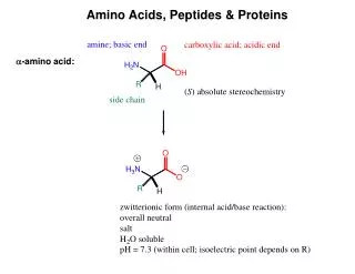
Amino acids • All proteins are composed of amino acids. • Twenty common amino acids. • All are -amino acids except proline. • A primary amine is attached to the carbon - the carbon just after the acid group. carbon H | R-C-COOH | NH2 General Structure
Amino acids • Because an acid and base are both present, an amino acid can form a +/- ion. • H H • | | • R-C-COOH R-C-COO- • | | • NH2 NH3+ • How well it happens is based on pH and the type of amino acid. Called azwitterion.
-Amino acids • Except for glycine, the carbon is attached to four different groups - chiral center. • Carbohydrates • We use the D-form. • Amino Acids • We use the L-form. COO- | H3N - C - H | R +
Classification of amino acids • The -amino acid group is the same in each. • Classified by the type of side chain. • Group I. non-polar side chains. • Group II. polar, uncharged side chains • Group III. acidic side chains • Group IV. basic side chains
Group I. Non-polar side chains H3CH \ | HC-C-COO- / | H3C+NH3 H | CH3-C-COO- | +NH3 alanine valine H3C H \ | HC-CH2-C-COO- / | H3C+NH3 H3CH || H3C-CH2-CH-C-COO- | +NH3 leucine isoleucine
H | -CH2-C-COO- | +NH3 H2C CH-COO- | | H2C+NH2 H2C H | CH2-C-COO- | +NH3 N H Group I. Non-polar side chains proline phenylalanine H | CH3 -S-CH2-CH2-C-COO- | +NH3 methionine tryptophan
H | -CH2-C-COO- | +NH3 HO- Group II. Polar side chains H | H-C-COO- | +NH3 H | HO-CH2-C-COO- | +NH3 glycine serine HO H | | CH3-CH-C-COO- | +NH3 tyrosine threonine
Group II. Polar side chains H | HS-CH2-C-COO- | +NH3 cysteine O H | || H2N-C-CH2-CH2-C-COO- | +NH3 O H | || H2N-C-CH2-C-COO- | +NH3 glutamine asparagine
Group III. Acidic side chains • Based on having a pH of 7. O H | || -O-C-CH2-CH2-C-COO- | +NH3 glutamic acid O H | || -O-C-CH2-C-COO- | +NH3 aspartic acid
Group IV. Basic side chains • Based on a pH of 7. H +| H3N-CH2-CH2-CH2-CH2-C-COO- | +NH3 lysine +NH2 H | || H2N-C-N-CH2-CH2-CH2-C-COO- | +NH3 H arginine H | CH2-C-COO- | +NH3 histidine H N H N H +
H | H2NCCOOH | R H | H2NCCOOH | R’ H O | || H2N - C - C - | R H | N - C - COOH | | H R’ + Polypeptides and proteins • Proteins are polymers made up of amino acids. • Peptide bond - how the amino acids are • linked together to make • a protein. + H2O
ala arg asn asp cys gln glu gly his ile leu lys met phe thr pro ser trp tyr val Polypeptides and proteins • Here is an example sequence of amino acids in a protein. • It also shows the abbreviations commonly used.
Polypeptides and proteins • Residue - term used to refer to the amino acid once incorporated into a polypeptide • Polypeptide - contain 10-100 residues • Protein - contain more than 100 residues. • Most peptides and proteins isolated from cells contain between 2 - 2000 residues. • An average amino acid has a weight of 110, so protein molecular weights are in the range of 220 - 220,000 (some are much larger).
H O | || H2N - C - C | R H O | || - NH - C - C - | R’ H | N - C - COOH | | H R’’ Peptides N-terminal residue C-terminal residue peptide linkages
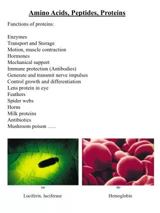
Protein function • Enzymes biological catalysts. • Immuno- antibodies of immune system. • globulins • Transport move materials around - • hemoglobin for O2. • Regulatory hormones, control metabolism. • Structural coverings and support - • skin, tendons, hair, nails, bone. • Movement muscles, cilia, flagella.
Protein size, composition and properties • One important property is molecular weight. There are two common ways to calculate it. • Determine the number of amino acid residues, then multiply by 110 -- the average molecular weight of an amino acid. • Directly measure the mass of a protein and report it in daltons. One dalton = One atomic mass unit.
Size of some important proteins • Protein MW Residues • Insulin 6,000 51 • Cytochrome c 16,000 104 • Hemoglobin 65,000 574 • Gamma globulin 176,000 1320 • Myosin 800,000 6100
Protein composition • Proteins can be classified based on the number of polypeptides used • Monomeric - only a single polypeptide chain is present. • Oligomeric - two or more polypeptide chains are present. • The subunit peptide chains are typically held together with noncovalent bonds.
Protein composition • Proteins are also classified based on their composition. • Simple proteins - only contain amino acid residues. • Conjugated proteins - contain other biomolecules - prosthetic groups. • These groups impart additional properties to a protein.
Example - cytochrome C 550 Heme structure Contains Fe2+ Used in metabolism. Aggregate of proteins and other structures.
Protein solubility • Two categories. • Determined by the types of amino acid side chains involved. • Water soluble • - globular proteins • Water insoluble • - fibrous proteins.
Four levels of protein structure • Primary structure • The actual sequence of amino acids in a protein. • Secondary structure • The type of regular repeating structure (-helix, -sheet) • Tertiary structure • Interaction of side chains. • Quaternary structure • Association of two or more polypeptide chains to form a multisubunit molecule.
H O | || H2N - C - C | R H O | || - NH - C - C - | R’ H | N - C - COOH | | H R’’ Summary ofprotein structure primary secondary tertiary quaternary
Determination of primary structure • The first step is to isolate the protein in a pure form from its natural source. Typically, only very small amounts can be obtained. • Total amino acid composition can be determined by hydrolysis of the protein. (6M HCl at 100oC). • The amount of each amino acid can then be measured chromatography.
Protein sequencing • Methods that determine the order of each amino acid in a protein. • Edman degradation. • Method of choice for protein sequencing. • Relies on a sequential degradation by removing one amino acid at a time from the N-terminus. • Process can be automated and works with peptides with up to 50 residues.
- NH COO 2 - COO - NH COO 2 H Edman degradation O O + CH C N CH C N R H R' phenylisothiocyanate peptide H S H O O N C N CH C N CH C N H R R' H+ isolate and react with additional reagent. O CH C N R' phenylthiohyantoin remaining peptide
Edman degradation • Problems with the method. • Does not provide 100% yield - resulting in contamination. • Limited to about 50 cycles so proteins must be cut to smaller sizes. Must rely on enzymes and reagents to cleave a protein at known locations. • Disulfide bonds between cysteine residues can present problems.
Protein sequencing • As of 1998, over 30,000 protein sequences were available in a computer database. • Having such information available makes it possible to study and compare sequence information. • Several biochemical conclusions have been made as a result of studying this data.
Protein sequencing • Identification of protein families. • Proteins with common sequence features have similar biological function, • This allow for the characterization of newly discovered proteins. • Example - protein kinases • Enzymes that catalyze the phosphorylation of amino acid residues. • All known protein kinases have the same common sequence region (domain) of 240 residues.
Protein sequencing • Evolutionary development of proteins • Comparison of protein types for many organisms. • Possible to establish taxonomic relationships. • Example - cytochrome c • Protein used in aerobic respiration. • It has been determined for over 60 organisms. • 27 residues are the same for all forms. • Other variations indicate evolutionary changes.
Protein sequencing • Search for dysfunction. • Normally, all residues in a protein are identical for a species. • Some individuals may produce a protein with one or more ‘incorrect’ residues. • Example - sickle cell anemia. • Two ‘incorrect’ amino acid residues result in malformed hemoglobin. • This causes deformation of red blood cells.
Protein sequencing • Three dimensional nature of proteins. • Sequence data can be coupled with other methods. • X-ray crystallography can produce 3-D structural information. • It is a difficult method and has not kept up with the number of proteins that have been isolated. • Sequencing may offer an alternative approach.
A l a A l a L y s P h e G l u A r g G l u H i s M e t A s p S e r S e r T h r S e r A l a A l a A l a S e r A s p T h r T h r S e r C y s T y r G l u S e r T y r S e r T h r M e t S e r G l u G l u I l e S e r G l y L y s C y s A l a T h r A s p N H H O O C - V a l 2 A s p L y s V a l S e r T r y A s p A s p A l a L y s C y s C y s A l a V a l C y s S e r G l u G l u A s p A s p A r g V a l A l a C y s G l u G l y A s p P r o T y r V a l P r o V a l H i s P h e V a l G l u G l u A s p I l e M e t T h r A l a I l e M e t G l y L e u L y s L y s S e r A s p S e r S e r A l a G l u T h r T h r L y s T y r A l a C y s A s p P r o T r y L y s S e r G l u H i s V a l P h e T h r A s p V a l P r o L y s C y s A r g A s p L y s T h r L e u A s p A r g Protein sequencing • Example - ribonuclease
Protein sequencing • Example - ribonuclease
Chromatography and electrophoresis of proteins • For a protein to be assayed by X-ray crystallography or protein sequencing, a pure sample must be produced. • After preparation of a cell extract, an appropriate separation method must be employed. Two such methods are: • Chromatography • Electrophoresis
Chromatography • Several chromatographic methods have been attempted to isolate pure protein fractions. • ion exchange • thin layer chromatography • column liquid chromatography • size exclusion chromatography • affinity chromatography • Affinity chromatography is becoming increasingly more important.
Affinity Chromatography • The method dates back to 1910. • Modern method was first published in 1967, by Axen, et al. -- ‘Cyanogen bromide Method for the Immobilization of Ligands on Agarose.’ • Ohlson (1978) was the first to demonstrate the use of a rigid, microparticulate support - beginnings of instrumental method.
Affinity Chromatography • The method involves the interaction of a ligand with the solute of interest. It can be viewed as being comparable to ion-exchange. • Two general types of ligands • Specific Binds only to one species. • Antibody/antigen • General Group specific • Binds to specific groups • on target species.
Affinity chromatography • Support • The material that the ligand is bound to. • Ideally, it should be rigid, stable and have a high surface area. • Agarose is the most popular although cellulose, dextran and polyacrylamide have been evaluated.
Affinity chromatography • Agarose gel • A polymer of D-galactose and 3,6-anhydro-L-galactose. • It can be used at pressures up to 1 psi and over a pH range of 4-9. • Cross-linking can be used to extend the pressure range.
Affinity chromatography • The separation is conducted in four basic steps. • Sample introduction • Adsorption of components of interest • Removal of impurities • Elution of components.
Affinity chromatography • Sample introduction • You must make sure • that your column has adequate capacity. ligand matrix spacer
Affinity chromatography • Absorption • Using a slow flow, your sample is then allowed to pass through the column. • The flow helps drive your sample components towards ‘fresh’ sites.
Affinity chromatography • Washing • Next, you can remove impurities by passing several volumes of fresh solvent through the column.
Affinity chromatography • Elution • The component of interest must then be removed and collected. • This also acts to regenerate the column.
Affinity chromatography • Elution methods • Biospecific • An inhibitor is added to the mobile phase (free ligand). • The free ligand will compete for the solute. • This approach is most often used when a low molecular weight inhibitor is available.
Affinity chromatography • Elution methods • Nonspecific • A reagent is added that denatures the solute (pH, KSCN, urea, ionic strength...) • Once denatured, the solute is released from the ligand. • If the solute is to be further used, it must not be irreversibly altered.
Affinity chromatography An example. Column: 50 mm x 30 mm containing 60 ml of Protein A Sepharose Sample: 5 liter cell culture supernatant with mouse IgGa2 and 0.5% fetal calf serum. Starting buffer: 0.1 M Na2HPO4, pH 7 Elution buffer: 0.1 M citric acid, pH 4 Flow rate: 66.6 ml/min 60 70 80 minutes
Electrophoresis • A separation method that relies on both the size and the charge of a species. • Samples are placed in an electrical field. • They tend to migrate to specific positions in the field. • With gel electrophoresis, a cross-linked polymer acts like a molecular sieve - smaller proteins move faster than larger ones.