Download
1 / 16
170 likes | 418 Views
Src Kinase Biosensor. Outline. Src Kinase Introduction Impacts of Src Src reporter components FPs (tECFP/EYFP) SH2 Flexible linker Substrate peptide 4. Fluorescent Proteins and FRET 5. Src Kinase Inactive and Active State

E N D
Src Kinase Biosensor
Outline • Src Kinase Introduction • Impacts of Src • Src reporter components • FPs (tECFP/EYFP) • SH2 • Flexible linker • Substrate peptide 4. Fluorescent Proteins and FRET 5. Src Kinase Inactive and Active State 6. How Src influence dynamical image of molecule in live cell 7. Linker, Substrate designation for a robust labeling protein
Introduction of Src Kinase • 1911 Peyton Rous isolated a virus from a chicken, which causes tumor in healthy bird, aka Rous sarcoma virus • V-src (sarcoma virus) consists of 3 simple genes: gag, pol, and envfor replication and encapsulation • 4th gene (v-src) codes for a protein which induces tumor cells. • C-src (cellular counterpart of v-src) affect signal transduction pathway to regulate cell-growth • Despite external signals, v-src activates internal control mechanism, hence induce oncogenic characterization.
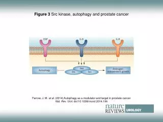
Impacts of Src activation • Impacts on cell polarity, adhesion, focal adhesion assembly/disassembly, lamellipodia formation, and migration. • Inhibition of Src results in impaired polarization toward migratory stimuli • Src phosphorylate cortactin. The phosphorylated cortactin associate and activate Arp2/3 to induce the growth of cortical actin network • Srcactivates the calpain-calpastatin proteolytic system to cleave FAK and disrupt focal adhesion complex => cell adhesion to ECM is reduced and cell motility is enhanced. • Src can phosphorylate p190RhoGAP and induce its binding to p120RasGAP => inhibition of RhoA, and subsequent dissolution of actin filaments.
Fluorescent Proteins and FRET • FPs: visualize signaling molecule • tECFP/EYFP pair • FRET: visualize dynamical molecular activities.
How does FRET work? • 2 chromophores are in proximity • Overlap of excitation spectrum of donor and acceptor • Energy transfer
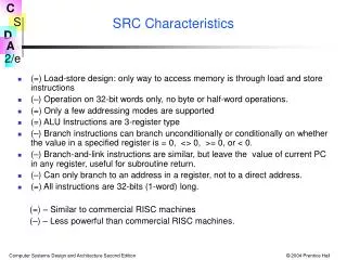
Src Kinase Structure • Non-receptor tyrosine kinases family • N-terminal SH4 domain • SH3 domain • SH2 domain (catalytic domain) • C-terminal regulatory sequence
How to activate Src Kinase? • Hormone binds cellular surface receptors (EGF, insulin) to generate phosphotyrosine • Phosphotyrosine attracts SH2 domain (homologous structure of src) to activate src.
Src inactive state vs. active state SH3 and SH2 couple, catalytic domain masked by C-terminal tail, prevent substrate binding. Interaction between integrin and Src changes Src conformation, thus activate it Integrin recruit RPTPα to dephosphorylate Y527 on C-terminal tail of Src and release it from kinase domain, thus make it active
FRET effect of Src reporter upon the actions of Src Kinase and Phosphatase
Emission Spectra of Src reporter before(Red) and after(black) phosphorylation by Src • When Src is inactivated, higher FRET is observed. • When Src is activated, emission intensity drops, thus yields lower FRET efficiency
Various Src biosensors with tECFP at N-termini and Citrine at C-termini