Download
1 / 74
740 likes | 755 Views
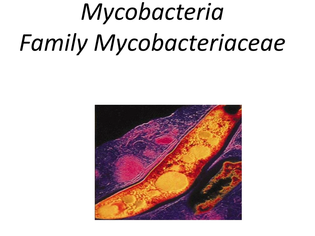
Mycobacteria Family Mycobacteriaceae. Mycobacteria. Slender bacilli which sometimes show branching, filamentous forms. A mould like pellicle growth in liquid culture media – therefore the name ‘mycobacteria ’ meaning ‘ fungus – like’ bacteria.

E N D
Mycobacteria • Slender bacilli which sometimes show branching, filamentous forms. • A mould like pellicle growth in liquid culture media – therefore the name ‘mycobacteria’ meaning ‘fungus –like’ bacteria. • Also called the ‘Acid Fast Bacilli’ or AFB because they are difficult to stain with ordinary dyes but once stained, resist decolourisation with mineral acids • Reason for acid fastness is the presence of ‘Mycolic acid’ in the cell wall
Classification of Mycobacteria Classification: Runyon 1959 classified the tuberculosis 1. Strict pathogens: (a). M.tuberculosiscomplex Closely antigenecally related, considered variants of same species. M. tb – human type M.bovis– bovine type M.africanum– human type M.microti– murine type
(b).Lepra bacilli: M. leprae – causing leprosy in man M.lepraemurium – causing rat leprosy (c). Other animal pathogens: M.microti – Murine type M.paratuberculosis – Johne’s bacillus.
2. Atypical mycobacteria: Group I – Photochromogeseg. M.kansasi Group II – Scotochromogenseg. M.scrofulaceum Group III – Non- chromogenseg. M.ulcerans Groups IV – Rapid growers eg. M.fortuitium 3. Saprophytic mycobacteria [non-pathogenic]: a. M.smegmatis – present in smegma, thermophiles. b. M.phlei – present in grass c. M.stercoris – Present in dung d. M.thermoresistible
Tuberculosis Definition–> A chronic granulomatous disease affecting humans and mammals. Etiology–> M.tuberculosis, M.bovis & M.africanum. • A Global emergency!!! • TB is an ancient infectious disease. • Tuberculosis kills > 5,000 people a day. • 2.3 million die each year. • 1/3 of world’s population is infected with TB. • 8 Million people develop active TB every year.
Bacterial virulence factors –> The ability of M.tuberculosisto survive & multiply within macrophages is responsible for: • initiation and spread of infection, • Persistence of infection & • Pathological tissue reactions observed in tuberculosis.
Cell wall lipids (Mycolic acids, Wax D, Phosphatides):- • Accounts for impermeability and resistance to antimicrobial agents • Resistance to attack by lysozyme, cationic proteins & oxygen radicals within phago-lysosome • Compounds including glycolipids, sulfatides down regulate the oxidative cytotoxic mechanism • resistance to osmotic lysis via complement deposition
Cord factor :- • The cord factor is primarily associated with virulent strains of Mycobacterium tuberculosis (Mtb). • Responsible for microscopic ‘serpentine cords’ wherein AFB are arranged in parallel chains in culture • It is known to be toxic to mammalian cells and to be an inhibitor of PMN migration, responsible for the chronic granulomatous reaction.
Host predisposing factors Acquiring infection –> large population, overcrowding, close contact and massive exposure. Natural resistance or susceptibility to M.tb–> dependent on the genetic set up of the individual -> higher incidence of disease has been suggested with the carriage of HLA-Bw15 antigen Age – Higher risk in infancy and in the elderly Nutritional status – severe malnutrition predisposes Underlying diseases – HIV infection, diabetes, other immuno-compromising disorders
Pathogenesis Source of infection -> • An open case of pulmonary tuberculosis. Bacilli are shed along with respiratory secretions while coughing, sneezing, to form “droplet nuclei.” Each droplet nuclei contains not less than 3 bacilli. Droplet nuclei are so small that they can remain air-borne for extended periods of time. The most effective (infective) droplet nuclei tend to have a diameter of 5 micrometers.
Less common sources :- Infected cow’s milk – Ingestion of unpasteurized milk from infected cows might result in acquiring infection. Clinical samples like sputum, fluids and cultures – inhalation of aerosols produced by manipulation of high positive sputum samples or cultures of M.tuberculosis
Mode of infection • Spread of droplet nuclei from one individual to another. • Tuberculosis begins when droplet nuclei reach the alveoli. • After droplet nuclei are inhaled, the bacteria are nonspecifically taken up by alveolar macrophages. • However, the macrophages are not activated and are unable to destroy the intracellular organisms.
Sequence of events Inhalation of tubercle bacilli <10% Adhesion by binding to complement and mannose receptors on surface of macrophag Phagocytosedby alveolar macrophages ( opsonization c3b mediated) Bacilli not killed due to bacterial cell wall LAM impairs phagosomelysosomefussion Multiply within phagocytes until they burst Bacilli kill macrophages, are taken up by migrating macrophages & spread to other body sites within such macrophages
The killing of macrophages now causes an acute inflammatory reaction. Edema fluid, lymphocytes, PMNL’s and later monocytes accumulating around the tubercle bacilli. This initial lesion is called the “Ghon’s focus” and this lesion along with associated regional lymph nodes is called the “Ghon’s complex”
By (3 – 8 weeks), an immune response is raised against the tubercle bacillus CMI is responsible for limiting active infection at this stage and most tubercle bacilli are killed by activated macrophages By activation of TH 1 & TH 2 Macrophage activating response Tissue damaging response IFN atcivate resting macrophages Intensified DTH response aggregate around the lessioncaseous necrosis becomes formation of tubercle liquified
The production & development of lesions and their healing or progression are determined by –> • The no. of mycobacteria in the inoculum and their subsequent multiplication • The resistance & hypersensitivity of the host
Types of Lesions • Exudative type:- The acute inflammatory reaction. Edema fluid,PMNLS& later monocytes around the tubercle bacilli Good CMI May lead to massive tissue necrosis May heal by resolution or, May develop into the productive type of lesion
Productive type:- This is a chronic granuloma, consisting of three zones –> • A central area of large, multinucleated giant cells containing the tubercle bacilli • A mid zone of pale epitheloid cells &, • A peripheral zone of fibroblasts, lymphocytes and monocytes
Later, peripheral fibrous tissue develops, and central area undergoes caseation necrosis – known as the “tubercle” A caseous tubercle may break into a bronchus, empty its contents there and form a cavity or, it may subsequently heal by fibrosis and calcification
Progression of Disease Tubercle bacilli spread within the host from the initial site by • Direct extension • Through the lymphatic channels & blood stream • Via the bronchi and the GIT During first infection, bacilli spread almost always via the lymphatics to regional lymph nodes –> blood stream –> to all organs (miliary distribution) Development of Miliary disease depends upon how much the host’s immune system is compromised.
Types of Tuberculosis • Primary tuberculosis:- • The first contact with the tubercle bacillus (usually during childhood in developing countries) • Acute exudative lesion develops and rapidly spreads to lymphatics & regional lymph nodes –> “Ghon’s Complex” • The lesion in tissue often heals rapidly • The lymph node usually undergoes massive caseation which later usually calcifies • Tuberculin test becomes positive
2. Reactivation tuberculosis or Post Primary tuberculosis:- • Caused by tubercle bacilli which had survived in the primary lesions • Almost always begins at the apex of lungs where O2 tension is highest • Characterized by chronic tissue lesions, formation of tubercles, fibrosis, caseation & shedding of tubercle bacilli in sputum • Extra pulmonary tuberculosis
Extra pulmonary Tuberculosis 1. Skeletal Tuberculosis: • Tuberculous osteomyelitis involves mainly the thoracic and lumbar vertebrae (Pott's disease) followed by knee and hip. • There is extensive necrosis and bony destruction with compressed fractures (with kyphosis) and extension to soft tissues, including psoas "cold" abscess
2. Genital Tract Tuberculosis: • In females, Tuberculous Salpingitis and granulomatous endometritis with irregular menstrual bleeding and infertility. • In males, tuberculosis involves prostate and epididymis most often with non-tender induration and infertility.
3. Urinary Tract Tuberculosis: • A "sterile pyuria" may suggest renal tuberculosis • Progressive destruction of renal parenchyma occurs if not treated. • Drainage to the ureters can lead to inflammation with ureteral stricture.
4. CNS Tuberculosis: • Tuberculous meningitis with CSF typically showing a high protein, low glucose, and lymphocytosis. • Rarely, a solitary granuloma, or "tuberculoma", may form and manifest with seizures. 5. Adrenal Tuberculosis: • Spread of tuberculosis to adrenals is usually bilateral, so that both adrenals are markedly enlarged. • Destruction of cortex leads to Addison's disease.
Gastrointestinal TB: • Uncommon today due to routine pasteurization of milk which eliminates M. bovis. • M.tbcoughed up in sputum may be swallowed into the GI tract. Lesions are circumferential ulcerations with stricture of the small intestine.
Scrofula: • Tuberculous lymphadenitis of cervical nodes may produce a mass of firm, matted nodes just under the mandible. • There can be chronic draining fistulous tracts to overlying skin. • This complication is usually seen in children
Symptoms of Pulmonary Tuberculosis • Cough (with/without phlegm) • Coughing up blood • Excessive sweating, especially at night • Fatigue • Fever • Weight loss • Breathing difficulty • Chest pain • Wheezing
LAB DIAGNOSIS: The current strategy is to look for novel approaches while also working for improvement in the existing diagnostics. • Direct and indirect methods help in laboratory diagnosis. • Demonstration of tubercle bacilli from clinical specimen and by culture is proof. - 50% pulmonary - 25% extra pulmonary tuberculosis + ve by smear taken from specimens.
Specimen collection:- • Pulmonary TB -> • Sputum • Induced sputum:Administration of inhaled bronchodilator and hypertonic saline. • Bronchoalveolar lavage (BAL) • Gastric lavage (in children)Nasopharyngeal aspiration,String test, • Sample collected by bronchoscopy
Extrapulmonary TB -> • CSF • Urine (early morning urine samples) • Pleural fluids • Joint fluids • Blood • Pus • Biopsy material
Direct Detection Methods MicroscopySputum smear microscopy still remains the basis for diagnosis of TB in developing countries. The most regular practice is Acid-fast staining using carbolfuschin and Fluorochromedye-auramine/rhodamine. Number of specimens As per the current Revised National Tuberculosis Control Programme (RNTCP) guidelines, the patient should visit the clinic at least twice to submit A spot – early morning or Spot-Spot Specimen.
80% of TB cases are detected with the first specimen. • 11.9% and 3.1% with the second and third specimen respectively. • Thus third sputum sample adds very little to the overall yield and omitting it will result in lesser patient visits to the clinic and decrease the lab workload, leading to lesser patient dropout and improved quality of service. Quantity and Quality of sputum: • At least 2 ml specimen should be collected. • which should be mucopurulent. • Sputum specimens should be examined within two days of collection.
Smear Microscopy Conventional method; Microscopy is the simplest and most rapid procedure currently availible to detect acid fast bacilli in clinical samples by; ZiehlNeelsen staining (hot method) Modifications: kinyoun method (cold method) Limit of detection at least 5x10³ bacilli per ml of sputum The result of smear can be influenced by Type of specimen Thickness of smear Extent of decolourisation UNCONCENTRATED SPUTUM
Advantage of smear microscopy: • It is inexpensive, simple. (ii) It is relatively easy to perform and read. (iii) Results can be reported within hours of receipt of the sample. (iv) Provides reliable epidemiological indicators needed for the evaluation of the National tuberculosis.
False + ve can occur in ZN staining; • Using same blotting paper to dry all smears. • Reusing slides. • Using the same dipping – decolorizing solutions for all smears may allow the floating free bacilli to stick to –ve smears. • Attached free bacilli on residual oil in the lens. • Use of tap water with saprophytes – usually acid fast only, uniform staining no beaded appearance is the difference.
Newer methods Chemical/ physical processing and concentration of sputum: Decontamination, Homogenization & Concentration:-
Petroff’s method Equal volumes of sample + 4% NaOH Incubated for 30 min with intermittent shaking Decontamination & homogenization takes place Centrifuged for 30 min at 1500 rpm Concentration of mycobacteria occurs Supernatant removed Neutralized with 8% HCl using Phenol red as indicator Concentrated sample microscopy/culture
Fluorescence microscopy : Smear microscopy with fluorochrome such as auramine-rhodamine. • Higher sensitivity while retaining similar specificity. • Less time consuming as smears can be examined at lower magnification. • Recent availability of LED light source for fluorescence microscopy, (earlier quartz-halogen lamps or high-pressure mercury vapour lamps were used) makes it useful for wide applicability. • RNTCP has adopted LED microscopy to replace ZN method in its designated microscopy centres (DMCs) across India.
Acid fast bacilli (AFB) within or outside pus cells. Fluorescent staining
Newer microscopic technologies Fluoresceindiacetate (FDA): Distinguish viable from dead bacilli • FDA stains only living, cultivable bacteria thus guiding antimicrobial therapy before culture reports come in. Automated microscopic technology by TBDx: Integrates robotic loading of stained slides and automated high-resolution digital image analysis to provide a result in minutes. CellScope: A portable digital FM that provides enlarged digitalised images for review.
Culture • Culture still remains the gold standard for diagnosis of TB. • Permits the diagnosis of drug resistance and the emerging mutations. • The limit of detection is 100 bacilli/ml, thus increasing the sensitivity compared to smear. Growth in a conventional egg-based medium takes anywhere from 4 to 8 weeks with an additional 4 weeks for drug sensitivity. Takes a median of 70 days to diagnose a case of MDR-TB by conventional culture methods
Processed specimen from non-sterile sites & centrifuged specimen from sterile sites. Inoculated onto various media, incubated at 37O C for up to 8 weeks. Media used
Colony characteristic • Inoculated media are examined daily for 4 days & then weekly up to 8 weeks. M.tb is identified by - • Slow growth (> 3 weeks) • Rough colonies with buff color • Acid fast staining shows AFB • Niacin & Nitrate reduction tests +ve
Liquid culture systems The most common detection method that is WHO endorsed in 2007, and adopted by the RNTCP, is a liquid culture using Middlebrook 7H9 broth – mycobacterial growth indicator tube (MGIT), A non-radiometric detection method which measures the consumption of oxygen by fluorescence