Download

1 / 43
450 likes | 668 Views
Transcription. MVA Fig. 26.8. Figure 31-11a X-Ray structure of Taq RNAP core enzyme. a subunits are yellow and green, b subunit is cyan, b¢ subunit is pink, w subunit is gray. What do you notice about this structure?. Page 1224.

E N D
Figure 31-11aX-Ray structure of Taq RNAP core enzyme. a subunits are yellow and green, b subunit is cyan, b¢ subunit is pink, w subunit is gray. What do you notice about this structure? Page 1224
Figure 31-13bModel of the open (Rpo) complex of Taq RNAP with promoter-containing DNA showing the transcription bubble and the active site. Page 1225
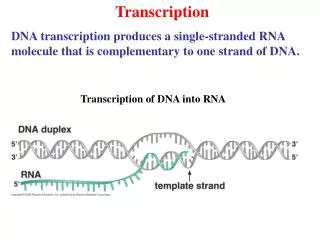
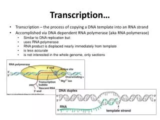
Figure 31-14 The two possible modes of RNA chain growth. Growth may occur (a) by the addition of nucleotides to the 3¢ end and (b) by the addition of nucleotides to the 5¢ end. Page 1226 How could you distinguish between these two possibilities?
DNA Footprinting http://users.rcn.com/jkimball.ma.ultranet/BiologyPages/F/ Footprinting.html
Figure 31-10 The sense (nontemplate) strand sequences of selected E. coli promoters. Page 1223
Figure 31-12a The sequence of a fork-junction promoter DNA fragment. Numbers are relative to the transcription start site, +1. Page 1225
Figure 31-15 RNA chain elongation by RNA polymerase. Page 1227
Figure 31-16 An electron micrograph of three contiguous ribosomal genes from oocytes of the salamander Pleurodeles waltl undergoing transcription. Page 1228
RNA Backtracking MVA Fig. 26.10
Figure 31-18 A hypothetical strong (efficient) E. coli terminator.
Regulation Possiblilites: Regulate transcription Regulate translation Regulate activity
The lac operon • E-coli uses three enzymes to take up and metabolize lactose. • The genes that code for these three enzymes are clustered on a single operon – the lac Operon. What’s lactose??
Figure 31-2 Genetic map of the E. coli lac operon. Page 1218
The lac repressor gene • Prior to these three genes is an operator region that is responsible for turning these genes on and off. • When there is not lactose, the gene for the lac repressor switches off the operon by binding to the operator region. • A bacterium’s prime source of food is glucose. • So if glucose and lactose are around, the bacterium wants to turn off lactose metabolism in favor of glucose metabolism.
Figure 31-25 The base sequence of the lac operator. Page 1239
Lac repressor binding to DNA animation http://molvis.sdsc.edu/atlas/morphs/lacrep/index.htm
Figure 31-28a X-Ray structures of CAP–cAMP complexes. (a) CAP–cAMP in complex with a palindromic 30-bp duplex DNA. Page 1241
Figure 31-36 X-Ray structure of the lac repressor subunit. Page 1248
Figure 31-37aX-ray structure of the lac repressor-DNA complex. Page 1249
Induction. • Allolactose is an isomer formed from lactose that derepresses the operon by inactivating the repressor, • Thus turning on the enzymes for lactose metabolism.
The lac operon in action. • When lactose is present, it acts as an inducer of the operon (turns it on). • It enters the cell and binds to the Lac repressor, causing a shape change that so the repressor falls off. • Now the RNA polymerase is free to move along the DNA and RNA can be made from the three genes. • Lactose can now be metabolized (broken down).
When the inducer (lactose) is removed • The repressor returns to its original shape and binds to the DNA, so that RNA polymerase can no longer get past the promoter. No RNA and no protein is made. • Note that RNA polymerase can still bind to the promoter though it is unable to move past it. That means that when the cell is ready to use the operon, RNA polymerase is already there and waiting to begin transcription.
Lac movie Lac and trp