Download

1 / 2
20 likes | 172 Views
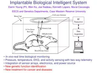
Bio-Complex-City. Telescope/Optical Transceiver. Light Beams. Van Rensselear Hall (33 rd St). Gbps Ethernet (fiber). Gbps Ethernet (fiber). Cellular Observatory Microscope System (Dr. J.Yasha Kresh et. al.). Remote observation and control (Dr. Banu Onaral et. al.).

E N D
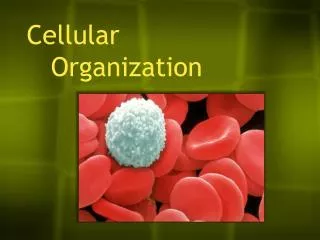
Bio-Complex-City Telescope/Optical Transceiver Light Beams Van Rensselear Hall (33rd St) Gbps Ethernet (fiber) Gbps Ethernet (fiber) Cellular ObservatoryMicroscope System (Dr. J.Yasha Kresh et. al.) Remote observation and control (Dr. Banu Onaral et. al.) Commonwealth Hall(31st St) MCP Hahnemann (15th St) COMPLEX BIOLOGICAL SYSTEMS RESEARCH P R O G R A M O V E R V I E W • The ability to observe and quantify mammalian live-cell phenomena is made possible largely because of advancements in visualization technology. In particular, cellular video microscopy (fluorescence, confocal), use of vital markers, and image-processing techniques have become the main stay of the modern cellular biology laboratory. A video microscopy-based 'collaboratory' has been developed to study cellular net-work dynamics, and in particular, to monitor live-cell spatio-temporal organization in real-time. The aim is to investigate the effects of intercellular communication on tissue genesis, differentiation, and cell survival. Cellular Network Dynamics: Monitoring Biological Organization in Real Time This virtual laboratory environment consists of cellular viability and survival studies conducted at Hahnemann University (Dr. J. Yasha Kresh, PI) and linked in real-time to image processing and modeling facilities at Drexel University (Dr. Banu Onaral, PI). Video monitoring of cellular culture in-vivo using immunofluorescence localization of inter- and intracellular network integrity (gap junctions, integrins, actin fibers) provides a source of live images collected over a span of minutes to hours. The ability to acquire a series of sequential images (e.g., beating myocytes, progression of cellular death), combined with advanced image enhancement, makes possible studies of cellular network dynamics (signaling complexity, self-organization). This ‘collaboratory,' made possible by high-speed net- work connectivity, creates opportunities for meaning- ful participation among researchers with diverse backgrounds, by facilitat- ing remote access to specialized equipment and dynamic processing or storage of vast amounts of complex data. This unique approach to distributed research has the advantages of greatly increasing the collective knowledge for distributed problem-solving projects. • Faculty/Contact: Y. Kresh, Ph.D., Research Director, Dept. of Cardiothoracic Surgery, Drexel U. College of Medicine; • B. Onaral, Ph.D., Director, School of Biomedical Engineering, Science & Health Systems, Drexel University. • E-mail: j.yasha.kresh@drexel.edu; banu.onaral@drexel.edu
Engineered Cellular Cultures Engineered 3-D construct for studying the topobiology of mechanically pre-programmed stem cells. Multi-electrode observatory of a self- organizing colony of cardiac cells. – Monitoring and control of cellular proliferation and differentiation. Example of myocardial tissue engineering and regeneration using adult stem cells. CELLULARNETWORK DYNAMICS: MONITORING BIOLOGICAL ORGANIZATION IN REAL TIME P R O J E C T O N E P A G E R • Engineered Cellular Cultures • Functional assays are being developed for observing and detecting the formation (making and breaking) of intercellular (junctional) communication in engineered tissue cultures, the assumption being that the relative increase in gap junction assembly is indicative of the integrative, 'cooperative' cellular networks (i.e., why form bonds or channels of communication do not do anything). The motivation for this line of thinking is related in part to the observation that the 'program' not to re-regenerate its cellular network structure limits the recovery of a damaged heart muscle. One approach in addressing this problem is to re-populate the impaired myocardium by healthy cells. In particular, our live-cell ‘observatory’ provides an ideal platform for studying the role that the extra-cellular microenvironment plays in the assimilation of non-cardiac cell types, such as fibroblasts, skeletal myoblasts, and adult stem (hematopoietic) cells. For example, observing the process of stem cell cardiomyocytes dynamic interaction (differentiation) in an ex-vivo setting may give clues for improving their integration within the intact cardiac cellular architecture. Cardiac function is mechanistically linked to junctional cell-cell contacts that give rise to synchronous electrical activity and coordinated mechanical contraction. In contrast, it has been observed that the ability of a tumor cell to proliferate is inversely related to the number of intercellular junctions that it forms. We are using the capabilities of the live-cell microscopy system to assess the functional attributes of the ensuing cardiac syncithium growing on electrically conductive polymers. Specifically, we will: a) visualize, quantitatively analyze, and model the topology of beating patterns; b)assess the establishment of intercellular connectivity, using lucifer yellow as a marker for cell-cell communication and; c) record the electrical activities using multi-array electrode chambers. Monitoring the cellular integration and tissue organization is a critical step in understanding the unfolding and emergence of organotypic function. The long-term objective of this research is to reverse-engineer biological principles and help inspire the design and engineering of robust and adaptive communication networks and intelligent systems. The advantages gained in mimicking biological organization and function are particularly intriguing. The cellular network models may have applicability in areas such as emergent communication networks, such as "building a network on a fly," evolutionary/adaptive optimization of networks, and distributed memory/smart agents. In addition, the biocomplexity inspired concepts of self-regulation and assembly can be explored 'in-silico' and generalized to study autonomous agent models, 2-D cellular automata, 'flocking' self-organized/coalition behavior, search pattern evolution problems, evolutionary optimization problems, as well as spatial genetic algorithms, among others. • Faculty/Contact:; Y. Kresh, Ph.D., Professor and Research Director, CT-Surgery, Drexel U. College of Medicine. • E-mail: j.yasha.kresh@drexel.edu • Collaborating Researchers: B. Onaral, Ph.D., Director, School of Biomedical Engineering, Science & Health Systems, Drexel University • Funding: Defense Advanced Research Projects Agency (DARPA); US Air Force Research Laboratory. • Laboratories: Cardiac Regeneration Laboratory; Molecular Cardiology Laboratory; Scaled Signals & Systems Laboratory.