Download
1 / 11
120 likes | 271 Views
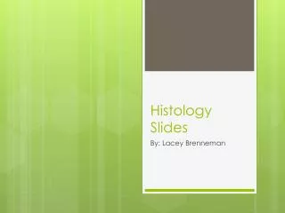
Histology Slides Part 2. Bio 210B. Motor Neuron. Motor Neurons. Motor Neuron. Spinal Cord ( c.s .). WM = white matter GM = gray matter.

E N D
Histology SlidesPart 2 Bio 210B
Spinal Cord (c.s.) WM = white matter GM = gray matter
Fig. 3a: Cross section through human spinal cord (lumbar region, L1) showing motor neurons. H&E method used. Astrocytes and oligodendrocytes are labeled 'A' and 'O', respectively. The objective magnification used is 40x.
Fig. 5a: Cross section through human spinal cord showing close-up view of internal structure of motor neuron located in the body of dorsal horn. Reduced silver method used. Silver grains are labeled 'S'. An oil immersion lens 100x has been used. Fig. 4: Cross section through human spinal cord showing motor neurons located in ventral horn. Reduced silver method used. Silver grains are labeled 'S'. The objective magnification used is 40x.
Blood Vessels, Nerve(s) and adipose Ignore the terms media & adventitia
a) Retina b) Photoreceptor layer (rods & cones) c) choiroid
Back of eye showing the optic disc and optic nerve, also visible are the layers of the cornea, the choroid coat and the sclera


![Read ebook [PDF] Histology Slides flash cards](https://cdn7.slideserve.com/12529330/slide1-dt.jpg)