Download

1 / 28
290 likes | 442 Views
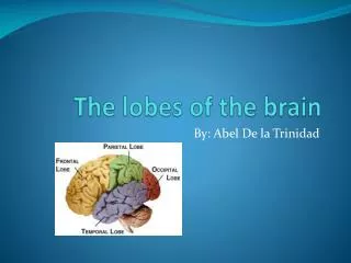
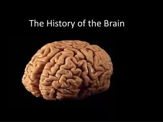
The History of the study of the Brain. Early brain studies used the method of phrenology . Phrenology – examination of the skull where bumps and indentations are used to make a ‘mental map’ and were then related to the character of that person.

E N D
The History of the study of the Brain • Early brain studies used the method of phrenology. • Phrenology – examination of the skull where bumps and indentations are used to make a ‘mental map’ and were then related to the character of that person. • The end of World War II and the massive quantity of brain injuries leads many people to examine the brain on a deeper level. • New technologies like the MRI have allowed doctors to observe the brain in operation in incredible detail. http://skepdic.com/phren.html
Overview – The Nervous System • Central Nervous System (CNS) = Brain + Spinal Cord • Peripheral Nervous System = Nerves to rest of body • Sensory nerves carry messages from the body to the brain • Motor nerves carry messages from the brain to the body
The Spinal Cord • The main pathway for information connecting the brain and peripheral nervous system - Conducts sensory information from the peripheral nervous system (both somatic and autonomic) to the brain. - Conducts motor information from the brain to the various effectors (skeletal muscles, cardiac muscle, smooth muscle, glands). • Serves as a minor coordinating center responsible for some simple reflexes (e.g., withdrawal reflex). • The average spinal cord is 45 cm long in men and 43 cm long in women. The spinal cord weighs approximately 35 g.
The Spinal Cord : Vertebrae • The human spinal cord is protected by the bony spinal column. The spinal column is made up of bones called vertebrae. • The spinal cord is located in the vertebral foramen and is made up of 30 segments: 7 cervical, 12 thoracic, 5 lumbar, 5 sacral and 1 coccyx. A pair of spinal nerves exits from each segment of the spinal cord.
Vertebra, thoracic (mid back) Vertebra, cervical (neck) Vertebra, lumbar (low back) Vertebral column
The vertebral column provides structural support for the trunk and surrounds and protects the spinal cord. • The vertebral column also provides attachment points for the muscles of the back and ribs. • The vertebral disks serve as shock absorbers during activities such as walking, running, and jumping. They also allow the spine to flex and extend. Common injury: Ruptured Disk
Complex lumbar spine fracture Cervical dislocation Thoracic dislocation Bullet in thoracic spinal canal
Famous Spinal Cord Injuries • Mike Utley – 1991 Detroit Lions Offensive Lion fractured his 6th and 7th cervical vertebrae. http://www.mikeutley.org/images/mike.gif
Cervical dislocation • Christopher Reeves • “Well, I totally decimated my first cervical vertebra and my second, and so my body and my spine and my head were not connected. Only my neck muscles were holding my head on, and fortunately I didn't suffer any brain damage, at least none that I can detect. [laughter] But, you know, that's what they tell me at any rate. But they literally had to put my head back on my body, and a wonderful surgeon, Dr. John Jane at the University of Virginia, was the one who operated on me, and they had to make it up. They had never done anything like this before, because this is what is called a hangman's injury, you know, like if you get dropped through the trap door and then cut down, sent to rehab and told to have a nice life.” http://record.wustl.edu/archive/2000/12-01-00/photos/reeves.jpg
Four ways the brain protects itself: • 1. Skull - • 2. Meninges • 3. Cerebrospinal Fluid • 4. Blood Brain Barrier (BBB)
Central Nervous System- The Coverings of the Brain (Shocks) Meninges • Both the brain and the spinal cord are covered in three continuous sheets of connective tissue, the meninges. • The dura mater (the dura): the outer layer • The arachnoid: the middle layer • The pia mater (the pia): the inner layer • “The meninges PAD the brain”
http://www.baltimoresun.com/media/photo/2004-05/12819309.jpg
Central Nervous System- Cerebrospinal Fluid (CSF) - The entire surface of central nervous system is bathed by a clear, colorless fluid, called cerebrospinal fluid (CSF). The CSF is contained within a system of fluid-filled cavities called ventricles. • The total volume of CSF is 125-150 ml • Normal resting pressure of the CSF is between 150-180 mmH2O. • Total production of CSF is about 400-500 ml/day (about 0.36 ml/min)
Central Nervous System- Cerebrospinal Fluid (CSF) - What does the CSF do? • Protection: The CSF protects the brain from damage by “buffering” the brain. • Buoyancy: The pressure at the base of the brain is reduced by immersing in the CSF. • Excretion of waste products: The one-way flow from the CSF to the blood takes potentially harmful metabolites, drugs and other substances away from the brain. • Endocrine medium for the brain: The CSF serves to transport hormones to other areas of the brain.
What is the Blood-Brain Barrier (BBB) ? The barrier exists between the blood and the brain
BBB – History • The late 19th century, Paul Ehrlich’s experimentCertain dyes (e.g., a series of aniline derivatives) administered intravenously to small animals stained all the organs except the brain: The brain has a lower affinity for the dye than the other tissues. In 1913, Edwin G. Goldman’s experiment The dye trypan blue, directly injected into the cerebrospinal fluid of rabbits or dogs, readily stained the entire brain but did not enter the bloodstream to stain the other internal organs: The CNS is separated from the blood by a barrier of some kind.
BBB – History • The late 1960’s Electron microscopy confirmed the hypothesis that brain capillaries provide the anatomical basis of the BBB • Recent studies - The BBB is present in all vertebrate brains - The BBB is laid down within the first 3 months of human fetal life
BBB – Function • Physical Barrier • To protect the brain tissues from “foreign substances” or “certain chemicals” in the blood that may injure the brain “Biological Shield” or “Safeguard”
BBB – Function“Neuroprotective Role”“Obstacle to Drug Delivery”
In order to cross the BBB in pharmacologically significant amounts, the therapeutic drugs must have the following characteristics: • Lipid soluble • Have a molecular weight < 400 Daltons • Not be a substrate for a BBB active efflux transporter This class of drugs constitutes <2% of all potential small molecule drugs. Only few brain disorders, such as depression, schizophrenia, chronic pain, and epilepsy, respond to such drugs. 100% of large molecule drugs and 98% of small molecule drugs do not cross the BBB.
Reflection • Of the four protective functions, which do you think would be most important in the field of psychology? Why?