Download
1 / 27
400 likes | 1.2k Views
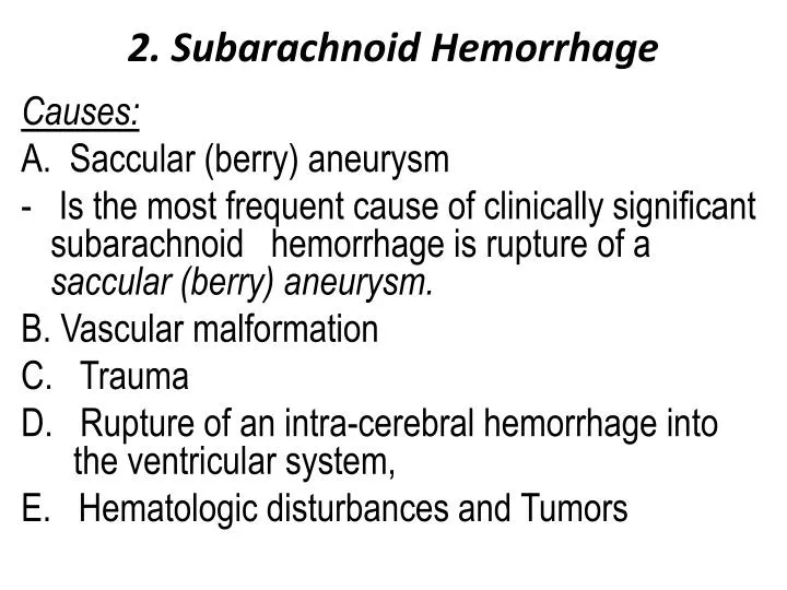
2. Subarachnoid Hemorrhage. Causes: A. Saccular (berry) aneurysm - Is the most frequent cause of clinically significant subarachnoid hemorrhage is rupture of a saccular (berry) aneurysm. B. Vascular malformation C. Trauma

E N D
2. Subarachnoid Hemorrhage Causes: A. Saccular (berry) aneurysm - Is the most frequent cause of clinically significant subarachnoid hemorrhage is rupture of a saccular (berry) aneurysm. B. Vascular malformation C. Trauma D. Rupture of an intra-cerebral hemorrhage into the ventricular system, E. Hematologic disturbances and Tumors
A. Saccular (Berry ) aneurysm: - Rupture of saccular aneurysm can occur at any time - In about one-third of cases it is associated with acute increases in intracranial pressure, such as with straining at stool or sexual orgasm. • The main symptom is sudden, excruciating headache (classically described as "the worst headache I've ever had") and rapidly lose consciousness. - Between 25% and 50% of individuals die with the first rupture, although those who survive typically
improve and recover consciousness in minutes - Recurring bleeding is common in survivors - It is currently not possible to predict which individuals will have recurrences of bleeding. - The prognosis worsens with each episode of bleeding. • About 90% of saccular aneurysms occur in the anterior circulation - Multiple aneurysms exist in 20% to 30% of cases.
Although they are referred to as congenital, they are not present at birth but develop over time because of underlying defects in the vessel media - It might be associated with: a. Disorders of extracellular matrix proteins, b. There is an increased risk of aneurysms in individuals with autosomal dominant polycystic kidney disease - Overall, aneurysms have a roughly 1.3% per year rate of bleeding.
- The probability of rupture increases with the size of the lesion, • Aneurysms greater than 10 mm have a roughly 50% risk of bleeding per year • Complications of subarachnoid hemorrhage 1- In the early period there is a risk of additional ischemic injury from vasospasm involving other vessels. 2- In the healing phase , meningeal fibrosis occurss sometimes leading to obstruction of CSF flow and interruption of pathways of CSF resorption.
Morphology - An un-ruptured saccular aneurysm is a thin-walled out-pouching of an artery. - The muscular wall are absent from the aneurysm sac itself - The sac is made up of thickened hyalinizedintima. - The adventitia covering the sac is continuous with that of the parent artery - Rupture usually occurs at the apex of the sac
other types of aneurysms present with cerebral infarction from vascular occlusion and include A. Atherosclerotic (fusiform) mostly of the basilar artery), B. Mycotic, traumatic, and dissecting aneurysms. , are most often found in the anterior circulation.
B. Vascular Malformations • Arteriovenous malformations (AVM) • Isthe most common type and the most dangerous - Affect males twice as frequently as females; • The lesion is most often recognized clinically between the ages of 10 and 30 years - The lesions presenting as : a. A seizure disorder b. An intra-cerebral hemorrhage c. A subarachnoid hemorrhage
Large AVMs occurring in the newborn period can lead to high-output congestive heart failure because of blood shunting directly from arteries to veins. Morphology - Involve vessels in the subarachnoid space extending into brain parenchyma or they may occur exclusively within the brain. Macroscopic appearance - They resemble a tangled network of wormlike vascular channels
- Microscopically • They are enlarged blood vessels separated by gliotic tissue, often with evidence of prior hemorrhage - Some vessels can be recognized as arteries with duplicated and fragmented internal elastic lamina, while others show marked thickening or partial replacement of the media by hyalinized connective tissue.
. 2. Cavernous hemangiomas - Consist of distended, loosely organized vascular channels with thin collagenized walls. - They are devoid of intervening nervous tissue - They occur most often in the cerebellum, pons, and subcortical regions, - Have a low flow without arterio-venous shunting. - Foci of old hemorrhage, infarction, and calcification frequently surround the abnormal vessels
3. Capillary telangiectasias - Are microscopic foci of dilated, thin-walled vascular channels separated by relatively normal brain parenchyma - Occurring most frequently in the pons. 4. Venous angiomas (varices) - Consist of aggregates of ectatic venous channels. Note: - The latter two types are unlikely to bleed and are most commonly discovered as incidental lesions.
. - Repetitive episodes of trauma (such as occurs in athletesparticipating in contact sports) can lead to later development of neurodegenerative processes a. Association of trauma with the risk of Alzheimer disease b. Chronic traumatic encephalopathy
I. Traumatic Parenchymal Injuries A. Contusions : - Results from blunt brain trauma - The pia-arachnoid is not breached - When an object impacts the head, brain injury may occur at the site of impact-a coup injury-or opposite the site of impact on the other side of the brain-a contrecoup injury. - Both coup and contrecoup lesions are contusions
1. Coup contusions - Occur at the site of impact in the absence of a fracture - Tend to be more severe in relation to a blow to the stationary head 2. Contrecoup contusions - Occur in the brain away from the point of impact and they occur more in relation to sudden deceleration of the head and the classic example is a fall on the occiput resulting in severe contusions to the frontal and temporal poles
Are caused by rapid tissue displacement, with subsequent hemorrhage and edema • The crests of the gyri are the most susceptible to traumatic injury. Than depth of sulci • Are common in regions of the brain overlying rough and irregular inner skull surfaces, such as: a. The frontal poles b. The orbital surfaces of the frontal lobes c. And the temporal lobe tips
.MORPHOLOGY : - Are wedge-shaped, and within a few hours of injury, blood extravasates across the the cerebral cortex, - In contrast with ischemic lesions, in which the superficial- layer of cortex may be preserved, trauma affects the superficial layers most severely. - Old contusions are are depressed, yellowish brown patches involving the crests of gyri -- More extensive regions of trauma give rise to large cavitary lesions, which resemble remote infarcts.
II. Diffuse axonal injury), • Trauma can also cause more subtle but widespread injury to axons within the brain with devastating consequences Mechanism: - The movement of one region of brain relative to another is thought to disrupt axonal integrity and function. - Angular acceleration, even in the absence of impact, may cause axonal injury as well as hemorrhage.
- As many as 50% of patients who develop coma shortly after trauma are believed to have white matter damage and diffuse axonal injury. - Although these injuries may be widespread, the lesions usually are asymmetric and are most commonly found a. Regions near the angles of the lateral ventricles .
b. Corpus callosum c. Various tracts in the brain stem -. They take the form of axonal swellings that appear within hours of the injury.
IV. Concussion - Describes reversible altered consciousness from head injury in the absence of contusion. -The characteristic transient neurologic dysfunction includes; 1. Loss of consciousness, 2. Temporary respiratory arrest 3. Loss of reflexes.
- Although neurologic recovery is complete, amnesia for the event persists. - The pathogenesis of the sudden disruption of nervous activity is unknown
V. Traumatic Vascular Injury . A. Epidural Hematoma - The middle meningeal artery is vulnerable to trauma - In infants, traumatic displacement of the easily deformable skull may tear a vessel, even in the absence of a skull fracture. - In children and adults, by contrast, tears involving dural vessels almost always stem from skull fractures -