Download
1 / 58
640 likes | 831 Views
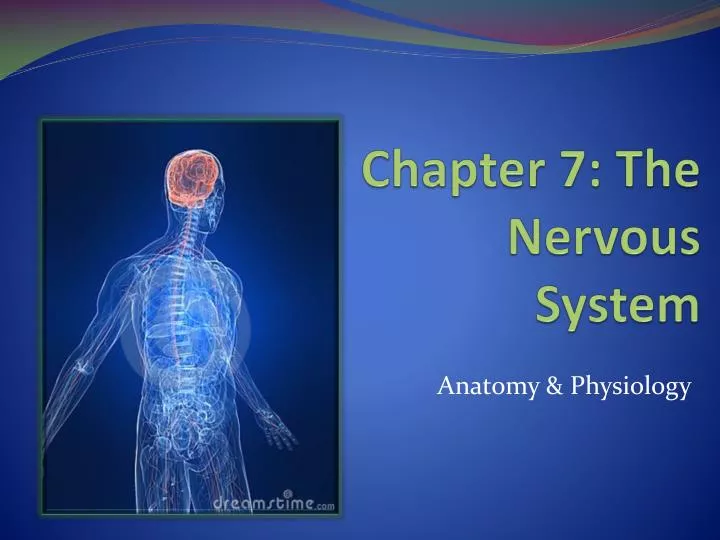
Chapter 7: The Nervous System. Anatomy & Physiology. Functions of the Nervous System. Nervous System manages body via electrical impulses Sensory input—gathering information Monitor changes occurring inside and outside the body Changes = stimuli Internal vs external Integration

E N D
Chapter 7: The Nervous System Anatomy & Physiology
Functions of the Nervous System • Nervous System manages body via electrical impulses • Sensory input—gathering information • Monitor changes occurring inside and outside the body • Changes = stimuli • Internal vs external • Integration • Processes and interprets sensory input • Decide if/what action is needed • Motor output • A response to integrated stimuli • By activating muscles or glands
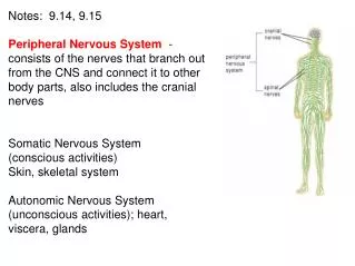
Structural Classification of the Nervous System • Central nervous system (CNS) • Brain • Spinal cord • Peripheral nervous system (PNS) • Nerves outside the brain and spinal cord • Spinal nerves • Cranial nerves
Classification of the Peripheral Nervous System • Sensory (afferent) division • Nerve fibers that carry information to the central nervous system • Via sensory fibers found in: • Skin • Skeletal muscles • Joints • Motor (efferent) division • Nerve fibers that carry impulses away from the central nervous system
Motor (Efferent) Division • Somatic nervous system = voluntary • System that we consciously control • I.E. contracting muscles on demand • Autonomic nervous system = involuntary • System that is activated subconsciously • I.E. cardiac muscles, glands, respiratory rate
Nervous Tissue: Support Cells • Astrocytes • Abundant, star-shaped cells • Brace neurons • Form barrier between capillaries and neurons • Control the chemical environment of the brain
Nervous Tissue: Support Cells • Microglia • Spiderlike phagocytes • Dispose of debris • i.e. dead brain cells • bacteria
Nervous Tissue: Support Cells • Ependymal cells • Line cavities of the brain and spinal cord • Circulate cerebrospinal fluid w/ cilia
Nervous Tissue: Support Cells • Oligodendrocytes • Wrap around nerve fibers in the central nervous system to form myelin sheaths • Schwann cells • Form myelin sheath in the peripheral nervous system • Include neurilemma. • Satellite cells • Protect neuron cell bodies
Nervous Tissue: Neurons • Neurons = nerve cells • transmit messages • Major regions of neurons • Cell body—nucleus and metabolic center of the cell • Processes—fibers that extend from the cell body • Cell body • Nissl substance • Specialized rough endoplasmic reticulum • Neurofibrils • Intermediate cytoskeleton • Maintains cell shape
Nervous Tissue: Neurons • Cell body • Nucleus • Large nucleolus • Projections outside the cell body • Dendrites—conduct impulses toward the cell body • Axons—conduct impulses away from the cell body
Nervous Tissue: Neurons • Axons end in axonal terminals • contain vesicles with neurotransmitters • Neurons don’t touch each other rather are separated by a gap • Synaptic cleft—gap between adjacent neurons • Synapse—junction between nerves
Nervous Tissue: Neurons • Myelin sheath—whitish, fatty material covering axons • increases speed of nerve impulses • Schwann cells—produce myelin sheaths in jelly roll–like fashion • Nodes of Ranvier—gaps in myelin sheath along the axon
Neuron Cell Body Location • Most neuron cell bodies are found in the central nervous system • Gray matter—cell bodies and unmyelinated fibers • Ganglia—collections of cell bodies outside the central nervous system • “Tracts” are nerve fibers traveling through the CNS • “Nerves” nerve fibers traveling through PNS
Functional Classification of Neurons • Sensory (afferent) neurons • Carry impulses from the sensory receptors to the CNS • Receptors • Cutaneous sense organs • Proprioceptors—detect stretch or tension in muscles, tendons, joints • Brain makes adjustments for posture and balance • Motor (efferent) neurons • Carry impulses from the central nervous system to viscera, muscles, or glands to illicit reaction • Interneurons (association neurons) • Found in neural pathways in the CNS • Connect sensory and motor neurons
Structural Classification of Neurons • Multipolar neurons —many extensions (processes) from the cell body • Common Location: Motor neurons
Structural Classification of Neurons • Bipolar neurons —one axon and one dendrite • Rare • Common locations: Special sense organs (i.e. ears, nose, eyes)
Structural Classification of Neurons • Unipolar neurons —have a short single process leaving the cell body • 1 process only • Axon: Includes CNS and PNS extension • Common Location: Sensory Neurons in PNS
Physiology of Nerve Impulses • Neurons function in 2 ways: • Irritability: Respond to stimulus and translate it into a nerve impulse • Conductivity: Transport impulse to necessary location (i.e. other neurons, muscles, glands, organs)
Physiology of Nerve Impulses • Membrane of Neuron • At rest: Membrane is impermeable to sodium ions 1)When activated: sodium ions rush into the neuron cell body. • How are they activitaed? • By external stimulus (i.e. light-eye, smell-nose, sound-ear) • Most commonly by neurotransmitters 2)Depolarization occurs (sodium-potassium pump: more sodium in than out) and results in a graded potential 3) If graded potential is strong enough it leads to…. • Action potential/nerve impulse
Physiology of Nerve Impulses 4) Repolarization: • Membrane becomes impermeable again • Sodium goes outside cell body, potassium goes inside • Restores natural state • Another impulse cannot be initiated until repolarizationoccurs • Uses what energy? • Myelin sheaths role: • Causes impulse to travel faster by… • Nerve impulse leaps OVER sheaths because it can’t travel through fat
Physiology of Nerve Impulses • How does nerve impulse travel throughout neurons? • Electrical chemical event: • Impulse cannot go past axon terminals • Vesicles release neurotransmitters • They travel across synaptic cleft • Bind to receptors on the dendrites of the next neuron • Initiate process of nerve impulse all over again https://www.youtube.com/results?search_query=nerve+impulse
Axonterminal Actionpotentialarrives Axon oftransmittingneuron Vesicles Synapticcleft Receivingneuron Synapse
The Reflex Arc • Reflex: A programmed, rapid, involuntary response to stimuli • Reflex arcs: Neural pathways that reflexes travel • 2 types of Reflexes: • Somatic: Reflexes involving skeletal muscle • i.e. removing hand from hot iron • Autonomic: Regulate smooth muscles organs, breathing rate, BP
The Reflex Arc • Reflex arcs made up of a minimum of 5 steps • Sensory Receptor • Sensory Neuron • Integration Center • Motor Neuron • Effector
Skin Spinal cord (in cross section) Stimulus at distalend of neuron Sensory neuron Integrationcenter Receptor Motor neuron Interneuron Effector (a)
Regions of the Brain • Cerebrum (Cerebral Hemispheres) • Diencephalon • Brain Stem • Cerebellum
Regions of the Brain • Cerebrum (Cerebral Hemispheres) • Paired (left & right) superior parts of the brain • Includes more than half of the brain mass • The surface is made of ridges (gyri) and grooves (sulci)
Regions of the Brain-CEREBRUM • Lobes of the cerebrum • Fissures (deep grooves) divide the cerebrum into lobes • Surface lobes of the cerebrum • Frontal lobe • Parietal lobe • Occipital lobe • Temporal lobe
Regions of the Brain-CEREBRUM • Specialized areas of the cerebrum • Primary somatic sensory area • Receives impulses from the body’s sensory receptors • (i.e. cold, pain, touch) • Located in parietal lobe • Primary motor area • Sends impulses to skeletal muscles • Located in frontal lobe • Broca’s Area • Involved in our area to speak
Regions of the Brain-CEREBRUM • Çerebral areas involved in special senses • Gustatory area (taste) • Location: Parietal lobe • Helps detect different tastes in foods • Visual area • Location: Occipital lobe • Recognition based on past experiences • Auditory area • Location: Temporal Lobe • Recognition/perception of sounds • Olfactory area • Location: Medial Temporal Lobe • Conscious awareness of scents
Regions of the Brain-CEREBRUM • Interpretation areas of the cerebrum • Located across both hemispheres (temporal, occipital, parietal lobes) • Speech/language region • Helps with understanding written/spoken language • Language comprehension region • Language/Reading comprehension • General interpretation area • Stores complex memories • Recognizes different senses/signals and puts them into a cognitive thought
Regions of the Brain-CEREBRUM • Layers of the cerebrum • Gray matter • outer layer in the cerebral cortex composed mostly of neuron cell bodies • White matter • fiber tracts deep to the gray matter • Corpus callosum connects hemispheres • Allows communication between them • Basal nuclei • sections of gray matter buried within the white matter • Adapts voluntary motor movements to changes
Regions of the Brain: Diencephalon • Sits on top of the brain stem • Enclosed by the cerebral hemispheres • Made of three parts • Thalamus • Hypothalamus • Epithalamus
Diencephalon THALAMUS HYPOTHALAMUS • FUNCTIONS: • Transports all sensory signals/impulses to the cerebral cortex (cerebrum) • Deals with sensations such as pain and temperature • FUNCTIONS: • Transports and receives signals/impulses from the cerebrum and thalamus • Works closely with the pituitary gland • Helps produce hormones to regulate activity of organs • Hunger • Metabolism • Body Temperature • Sleep • Emotions
Diencephalon • EPITHALAMUS • Houses the pineal body (an endocrine gland) • Includes the choroid plexus—forms cerebrospinal fluid
Regions of the Brain: Brain Stem • Attaches to the spinal cord • Parts of the brain stem • Midbrain • Pons • Medulla oblongata
Regions of the Brain: Brain Stem MIDBRAIN PONS • Location: • Between pons and diencephalon • Nerve fibers join brain stem and spinal cord to cerebrum • Functions: • Controls vision and hearing • Rounded bulge on brain stem • Between midbrain and medulla oblongata • Functions: • Control swallowing CN: V • Helps in facial movement CN VII
Regions of the Brain: Brain Stem • MEDULLA OBLONGATA • LOCATION: • Between pons and spinal cord • Functions: • Receives and interprets signals from spinal cord • Controls vital signs • Respiration (breathing rate) • Circulation • Blood pressure • Coughing • Heart beat (Beats per minute)
Regions of the Brain: Brain Stem • Reticular Formation • Diffuse mass of gray matter along the brain stem • Plays a role in awake/sleep cycles and consciousness
Regions of the Brain: CEREBELUM • Cerebellum a.k.a. “little brain” • Location: Attaches to brainstem and behind pons • Functions: • Center for balance • Motor control • Activates receptors in muscles, tendons, and joints
Protection of the Central Nervous System • Structures that offer protection: • Scalp and skin • Skull and vertebral column • Meninges • Cerebrospinal fluid (CSF) • Blood-brain barrier