Download
1 / 46
510 likes | 908 Views
Heart Failure. S. Soliman MD. Definition:. A state in which the heart cannot provide sufficient cardiac output to satisfy the metabolic needs of the body It is commonly termed congestive heart failure (CHF) since symptoms of increase venous pressure are often prominent. Etiology .

E N D
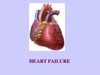
Heart Failure S. Soliman MD
Definition: • A state in which the heart cannot provide sufficient cardiac output to satisfy the metabolic needs of the body • It is commonly termed congestive heart failure (CHF) since symptoms of increase venous pressure are often prominent
Etiology • It is a common end point for many diseases of cardiovascular system • It can be caused by : -Inappropriate work load (volume or pressure overload) -Restricted filling -Myocyte loss
Causes of left ventricular failure • Volume over load: Regurgitate valve High output status • Pressure overload:Systemic hypertension Outflow obstruction • Loss of muscles:Post MI, Chronic ischemia Connective tissue diseases Infection, Poisons (alcohol,cobalt,Doxorubicin) • Restricted Filling:Pericardial diseases, Restrictive cardiomyopathy, tachyarrhythmia
Pathophysiology • Hemodynamic changes • Neurohormonal changes • Cellular changes
Hemodynamic changes • From hemodynamic stand point HF can be secondary to systolic dysfunction or diastolic dysfunction
Cellular changes Changes in Ca+2 handling. Changes in adrenergic receptors: •Slight in α1 receptors • β1 receptors desensitization followed by down regulation Changes in contractile proteins Program cell death (Apoptosis) Increase amount of fibrous tissue
Symptoms • SOB, Orthopnea, paroxysmal nocturnal dyspnea • Low cardiac output symptoms • Abdominal symptoms:Anorexia,nausea, abdominal fullness, Rt hypochondrial pain
Physical Signs • High diastolic BP & occasional decrease in systolic BP (decapitated BP) • JVD • Rales(Inspiratory) • Displaced and sustained apical impulses • Third heart sound – low pitched sound that is heard during rapid filling of ventricle
Physical signs (cont.) • Mechanism of S3 sudden deceleration of blood as elastic limits of the ventricles are reached • Vibration of the ventricular wall by blood filling • Common in children
Physical signs (cont.) • Fourth heart Sound (S4) - Usually at the end of diastole - Exact mechanism is not known Could be due to contraction of atrium against stiff ventricle • Pale, cold sweaty skin
Framingham Criteria for Dx of Heart Failure • Major Criteria: • PND • JVD • Rales • Cardiomegaly • Acute Pulmonary Edema • S3 Gallop • Positive hepatic Jugular reflex • ↑ venous pressure > 16 cm H2O
Dx of Heart Failure (cont.) • Minor Criteria LL edema, Night cough Dyspnea on exertion Hepatomegaly Pleural effusion ↓ vital capacity by 1/3 of normal Tachycardia 120 bpm Weight loss 4.5 kg over 5 days management
Forms of Heart Failure • Systolic & Diastolic • High Output Failure • Pregnancy, anemia, thyrotoxisis, A/V fistula, Beriberi, Pagets disease • Low Output Failure • Acute • large MI, aortic valve dysfunction--- • Chronic
Forms of heart failure ( cont.) • Right vs Left sided heart failure: Right sided heart failure : Most common cause is left sided failure Other causes included : Pulmonary embolisms Other causes of pulmonary htn. RV infarction MS Usually presents with: LL edema, ascites hepatic congestion cardiac cirrhosis (on the long run)
Differential diagnosis • Pericardial diseases • Liver diseases • Nephrotic syndrome • Protein losing enteropathy
Laboratory Findings • Anemia • Hyperthyroid • Chronic renal insuffiency, electrolytes abnormality • Pre-renal azotemia • Hemochromatosis
Electrocardiogram • Old MI or recent MI • Arrhythmia • Some forms of Cardiomyopathy are tachycardia related • LBBB→may help in management
Chest X-ray • Size and shape of heart • Evidence of pulmonary venous congestion (dilated or upper lobe veins → perivascular edema) • Pleural effusion
Echocardiogram • Function of both ventricles • Wall motion abnormality that may signify CAD • Valvular abnormality • Intra-cardiac shunts
Cardiac Catheterization • When CAD or valvular is suspected • If heart transplant is indicated
TREATMENT • Correction of reversible causes • Ischemia • Valvular heart disease • Thyrotoxicosis and other high output status • Shunts • Arrhythmia • A fib, flutter, PJRT • Medications • Ca channel blockers, some antiarrhythmics
Diet and Activity • Salt restriction • Fluid restriction • Daily weight (tailor therapy) • Gradual exertion programs
Diuretic Therapy • The most effective symptomatic relief • Mild symptoms • HCTZ • Chlorthalidone • Metolazone • Block Na reabsorbtion in loop of henle and distal convoluted tubules • Thiazides are ineffective with GFR < 30 --/min
Diuretics (cont.) • Side Effects • Pre-renal azotemia • Skin rashes • Neutropenia • Thrombocytopenia • Hyperglycemia • ↑ Uric Acid • Hepatic dysfunction
Diuretics (cont.) • More severe heart failure → loop diuretics • Lasix (20 – 320 mg QD), Furosemide • Bumex (Bumetanide 1-8mg) • Torsemide (20-200mg) Mechanism ofaction: Inhibit chloride reabsortion in ascending limb of loop of Henle results in natriuresis, kaliuresis and metabolic alkalosis Adverse reaction: pre-renal azotemia Hypokalemia Skin rash ototoxicity
K+ Sparing Agents • Triamterene & amiloride – acts on distal tubules to ↓ K secretion • Spironolactone (Aldosterone inhibitor) recent evidence suggests that it may improve survival in CHF patients due to the effect on renin-angiotensin-aldosterone system with subsequent effect on myocardial remodeling and fibrosis
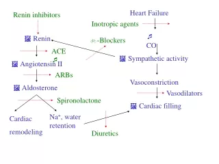
Inhibitors of renin-angiotensin- aldosterone system • Renin-angiotensin-aldosterone system is activation early in the course of heart failure and plays an important rolein the progression of the syndrome • Angiotensin converting enzyme inhibitors • Angiotensin receptors blockers • Spironolactone
Angiotensin Converting Enzyme Inhibitors • They block the R-A-A system by inhibiting the conversion of angiotensin I to angiotensin II → vasodilation and ↓ Na retention • ↓ Bradykinin degradation ↑ its level → ↑ PG secretion & nitric oxide • Ace Inhibitors were found to improve survival in CHF patients • Delay onset & progression of HF in pts with asymptomatic LV dysfunction • ↓ cardiac remodeling
Side effects of ACE inhibitors • Angioedema • Hypotension • Renal insuffiency • Rash • cough
Angiotensin II receptor blockers • Has comparable effect to ACE I • Can be used in certain conditions when ACE I are contraindicated (angioneurotic edema, cough)
Digitalis Glycosides (Digoxin, Digitoxin) • The role of digitalis has declined somewhat because of safety concern • Recent studies have shown that digitals does not affect mortality in CHF patients but causes significant • Reduction in hospitalization • Reduction in symptoms of HF
Digitalis (cont.)Mechanism of Action • +ve inotropic effect by ↑ intracellular Ca & enhancing actin-myosin cross bride formation (binds to the Na-K ATPase → inhibits Na pump → ↑ intracellular Na → ↑ Na-Ca exchange • Vagotonic effect • Arrhythmogenic effect
Digitalis Toxicity • Narrow therapeutic to toxic ratio • Non cardiac manifestations Anorexia, Nausea, vomiting, Headache, Xanthopsia sotoma, Disorientation
Digitalis Toxicity • Cardiac manifestations • Sinus bradycardia and arrest • A/V block (usually 2nd degree) • Atrial tachycardia with A/V Block • Development of junctional rhythm in patients with a fib • PVC’s, VT/ V fib (bi-directional VT)
Digitalis ToxicityTreatment • Hold the medications • Observation • In case of A/V block or severe bradycardia → atropine followed by temporary PM if needed • In life threatening arrhythmia → digoxin-specific fab antibodies • Lidocaine and phenytoin could be used – try to avoid D/C cardioversion in non life threatening arrhythmia
β Blockers • Has been traditionally contraindicated in pts with CHF • Now they are the main stay in treatment on CHF & may be the only medication that shows substantial improvement in LV function • In addition to improved LV function multiple studies show improved survival • The only contraindication is severe decompensated CHF
Vasodilators • Reduction of afterload by arteriolar vasodilatation (hydralazin) reduce LVEDP, O2 consumption,improve myocardial perfusion, stroke volume and COP • Reduction of preload Byvenous dilation ( Nitrate)↓ the venous return ↓ the load on both ventricles. • Usually the maximum benefit is achieved by using agents with both action.
Positive inotropic agents • These are the drugs that improve myocardial contractility (β adrenergic agonists, dopaminergic agents, phosphodiesterase inhibitors), dopamine, dobutamine, milrinone, amrinone • Several studies showed ↑ mortality with oral inotropic agents • So the only use for them now is in acute sittings as cardiogenic shock
Anticoagulation (coumadine) • Atrial fibrillation • H/o embolic episodes • Left ventricular apical thrombus
Antiarrhythmics • Most common cause of SCD in these patients is ventricular tachyarrhythmia • Patients with h/o sustained VT or SCD → ICD implant
Antiarrhythmics (cont.) • Patients with non sustained ventricular tachycardia • Correction of electrolytes and acid base imbalance • In patients with ischemic cardiomyopathy → ICD implant is the option after r/o acute ischemia as the cause • In patients wit non ischemic cardiomyopathy management is ICD implantation
New Methods • Implantable ventricular assist devices • Biventricular pacing (only in patient with LBBB & CHF) • Artificial Heart
Cardiac Transplant • It has become more widely used since the advances in immunosuppressive treatment • Survival rate • 1 year 80% - 90% • 5 years 70%
Prognosis • Annual mortality rate depends on patients symptoms and LV function • 5% in patients with mild symptoms and mild ↓ in LV function • 30% to 50% in patient with advances LV dysfunction and severe symptoms • 40% – 50% of death is due to SCD