Download
1 / 40
690 likes | 2.09k Views
Airway intubation. Joana Almeida. Anatomy. Methods:. Endotracheal intubation Orotracheal Nasotracheal Cricothyrotomy Tracheotomy. Endotracheal Intubation. Placement of a flexible plastic tube into the trachea to: maintain an open airway,

E N D
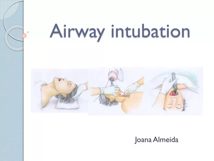
Airway intubation Joana Almeida
Methods: • Endotracheal intubation • Orotracheal • Nasotracheal • Cricothyrotomy • Tracheotomy
Endotracheal Intubation • Placement of a flexible plastic tube into the trachea to: • maintain an open airway, • serve as a conduit through which to administer certain drugs. • Is performed in critically injured, ill or anesthetized patients: • to facilitate ventilation of the lungs, including mechanical ventilation, • to prevent the possibility of asphyxiation or airway obstruction.
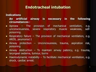
Indications: • For supporting ventilation in patient with pathologic disease: • Upper airway obstruction, • Respiratory failure, • Loss of consciousness • For supporting ventilation during general anaesthesia: • Type of surgery: • Operative site near the airway, • Thoracic or abdominal surgery, • Prone or lateral surgery, • Long period of surgery • Patient has risk of pulmonary aspiration • Difficult mask ventilation
Airway Assessment • 1) Condition that associated with difficult intubation: • Congenital anomalies Pierre Robin syndrome , Down’s syndrome • Infection in airway Retropharyngeal abscess, Epiglottitis • Tumor in oral cavity or larynx • Enlarge thyroid glandtrachea shift to lateral or compressedtracheal lumen
Continuation... • Maxillofacial ,cervical or laryngeal trauma • Temperomandibular joint dysfunction • Burn scar at face and neck • Morbidly obese or pregnancy
Airway Assessment • 2)Interincisor gap : normal more than 3 cms
Soft palate Uvula • 3)Mallampati classification: Class 3,4may be difficult intubation
Laryngoscopic view • Grade 3,4 risk for difficult intubation!
4) Thyromental distance : more than 6 cms • 5) Flexion and extension of neck
Preparing the procedure... Essentials that must be present to ensure a safe intubation! They can be remembered by the mnemonic SALT • Suction. This is extremely important. Often patients will have material in the pharynx, making visualization of the vocal cords difficult. • Airway. the oral airway is a device that lifts the tongue off the posterior pharynx, often making it easier to mask ventilate a patient. The inability to ventilate a patient is bad. Also a source of O2 with a delivery mechanism (ambu-bag and mask) must be available. • Laryngoscope. This lighted tool is vital to placing an endotracheal tube. • Tube. Endotracheal tubes come in many sizes. In the average adult a size 7.0 or 8.0 oral endotracheal tube will work just fine.
Miller blade Macintosh blade LARYNGOSCOPIC BLADE • Macintosh (curved) and Miller (straight) blade • Adult : Macintosh blade, small children : Miller blade
High volume Low pressure cuff Low volume High pressure cuff • Endotracheal tube cuff
Instruments used... • Self-refilling bag-valve combination (eg, Ambu bag) or bag-valve unit (Ayres bag), connector, tubing, and oxygen source. Assemble all items before attempting intubation. 2. Tincture of benzoin and precut tape. 3. Introducer (stylets or Magill forceps). 4. Suction apparatus (tonsil tip and catheter suction). 5. Syringe, 10-mL, to inflate the cuff. 6. Mucosal anesthetics (eg, 2% lidocaine) 7. Water-soluble sterile lubricant. 8. Gloves.
Tecnique: Sniffing position Flexion at lower cervical spine Extension at atlanto-occipital joint
Topical Anesthesia: Anesthetize the mucosa of the oropharynx, and upper airway withlidocaine 2%,if time permits and the patient is awake. • Direct Laryngoscopy: • Place the patient in the sniffing position. • 2. Check the laryngoscope and blade for proper fit, and make sure that the light works. • 3. Make sure that all materials are assembled and close at hand. MADgicWand™ Mucosal Atomization Device for atomizing topical solutions. With 5mL syringe
Curved blade technique • Open the patient's mouth with the right hand, and remove any dentures. • Grasp the laryngoscope in the left hand • Spread the patient's lips, and insert the blade between the teeth, being careful not to break a tooth. • Pass the blade to the right of the tongue, and advance the blade into the hypopharynx, pushing the tongue to the left. • Lift the laryngoscope upward and forward, without changing the angle of the blade, to expose the vocal cords.
Curvedblade technique • The anesthesiologist then takes the endotracheal tube, made of flexible plastic, in the right hand and starts inserting it through the mouth opening. • The tube is inserted through the cords to the point that the cuff rests just below the cords • Finally, the cuff is inflated to provide a minimal leak when the bag is squeezed Using a stethoscope , the anesthesiologist listens for breathing sounds to ensure correct placement of the tube
Straight blade technique Follow the steps outlined for curved blade technique, but advance the blade down the hypopharynx, and lift the epiglottis with the tip of the blade to expose the vocal cords. The tip of the laryngoscope blade fits below the epiglottis, which is no longer visible with the blade in position.
Video: • http://www.youtube.com/watch?v=tKz2zadEX_0&feature=related
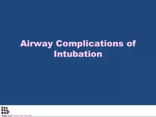
Complications: • Tube malpositioning(esophageal intubation ) • Tube malfunction or physiologic responses to airway instrumentation • Trauma such as tooth damage, lip/tongue/mucosal laceration, sore throat, dislocated mandible • Mucosal inflammation and ulceration and excoriation of nose can occur while the tube is in place • Laryngeal malfunction and aspiration, glottic, subglottic or tracheal edema and stenosis, vocal cord granuloma or paralysis during extubation • Physiologic responses to intubation include hypertension, tachycardia, intracranial hypertension, and laryngospasm
Laryngeal Masks(LMA) The Laryngeal Mask Airway is an alternative airway device used for anesthesia and airway support. • They cause less pain and coughing than an endotracheal tube, and are much easier to insert. It consists of an inflatable silicone mask and rubber connecting tube. It is inserted blindly into the pharynx, forming a low-pressure seal around the laryngeal inlet and permitting gentle positivepressure ventilation. All parts are latex-free
Laryngeal Masks • Short Procedure: • The cuff of the mask is deflated before insertion and lubricated. • The patient is sedated or fully anaesthetized if conscious, and their neck is extended and their mouth opened widely. • The apex of the mask, with its open end pointing downwardstoward the tongue, is pushed backwards towards the uvula. • The cuff follows the natural bend of the oropharynx, and its long walls come to rest over the piriform fossa. • Once placed, the cuff around the mask is inflated with air to create a tight seal. Indications: When endotracheal intubation is not necessaryor it’s difficult • Contraindications: • Non-fasted patients • Morbidly obese patients • Obstructive or abnormal lesions of the oropharynx Air entry is confirmed by listening for air entry into the lungs with a stethoscope
Advantages vs. Disadvantages • Advantages: • Allows rapid access • Does not require laryngoscope • Relaxants not needed • Provides airway for spontaneous or controlled • ventilation • Tolerated at lighter anesthetic planes • Disadvantages: • Does not fully protect against aspiration in the non-fasted patient • Requires re-sterilization
Nasoendotracheal intubation • Advantages: • 1) Comfortable for prolong intubation in postoperative period • 2) Suitable for oral surgery : tonsillectomy , mandible surgery • 3) For blind nasal intubation • 4) Can take oral feeding • 5) Resist for kinking and difficult to accidental extubation • Disadvantages: • 1) Trauma to nasal mucosa • 2) Risk for sinusitis in prolong intubation • 3) Risk for bacteremia • 4) Smaller diameter than oral route difficult for suction
Contraindication for nasoendotracheal intubation • 1) Fracture base of skull • 2) Coagulopathy • 3) Nasal cavity obstruction • 4) Retropharyngeal abscess
Complication of endotracheal intubation • 1) During intubation • 2) During remained intubation • 3) During extubation • 4) After extubation
1) During intubation • Trauma to lip, tongue or teeth • Hypertension and tachycardia or arrhythmia • Pulmonary aspiration • Laryngospasm • Bronchospasm • Laryngeal edema • Arytenoid dislocation hoarseness • Increased intracranial pressure • Spinal cord trauma in cervical spine injury • Esophageal intubation
2) During remained intubation • Obstruction from klinking , secretion or overinflation of cuff • Accidental extubation or endobronchial intubation • Disconnection from breathing circuit • Pulmonary aspiration • Lib or nasal ulcer in case with prolong period of intubation • Sinusitis or otitis in case with prolong nasoendotracheal intubation 3) During Extubation • Laryngospasm • Pulmonary aspiration • Edema of upper airway
4)After Extubation • Sore throat • Hoarseness • Tracheal stenosis (Prolong intubation) • Laryngeal granuloma
Cricothyrotomy • Incision made through the skin and cricothyroid membrane to establish a patent airway during certain life-threatening situations, such as airway obstruction by a foreign body, angioedema, or massive facial trauma. • Is easier and quicker to perform than tracheotomy, does not require manipulation of the cervical spine and is associated with fewer complications. • Used almost exclusively in emergency circumstances
Tracheotomy • Making an incision on the front of the neck and opening a direct airway through an incision in the trachea. • Allows a person to breathe without the use of their nose or mouth • Used primarily in situations where a prolonged need for airway support is anticipated.