Download
1 / 25
480 likes | 2.41k Views
Thrombotic Thrombocytopenic Purpura. History. In 1924, Dr.Eli Moschcowitz described a 16- year old girl with abrupt onset of petechiae, pallor, followed by paralysis, coma, and death. Autopsy showed ‘hyaline’ thrombi occluding terminal arterioles and capillaries. Definition.

E N D
History • In 1924, Dr.Eli Moschcowitz described a 16- year old girl with abrupt onset of petechiae, pallor, followed by paralysis, coma, and death. • Autopsy showed ‘hyaline’ thrombi occluding terminal arterioles and capillaries. British J of Hematology 2000
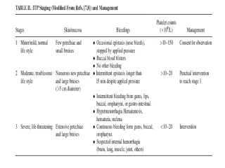
Definition • Syndrome of Coomb’s negative microangiopathic hemolysis and thrombocytopenia in the absence of an alternative explanation for these manifestations. • Presence of Fever, Neurological and renal abnormalities : classic Pentad. Rock GA, Br J Hematology 2000
Clinical Presentation • Approximately 1000 new cases occur each year • Common in middle aged group,median age-40 • Female:male (2:1). • Acute onset and fulminant course • Mortality rate >90% in pre-pheresis era. • Relapse rates, 10-40% ranging from months to years have been reported. Shumak KH, Ann Intern Med 1995
Clinical Features • Renal abnormalities • Proteinuria/hematuria > oliguria/ARF • Neurological abnormalities • Mental status changes> focal abnormalities. • Profound weakness related to anemia. • Abdominal pain,nausea, vomiting & diarrhea • Fever without chills. • Primary/idiopathic TTP vs. secondary
Secondary TTP • Drug-induced • Acute immune mediated: Ticlopidine & plavix. • Dose-related: mitomycin, tacrolimus, pencillin, cyclosporine, cisplatin, bleomycin, OCP • Quinine: HUS like illness. • Pregnancy and post-partum. • Allogenic bone marrow transplant. • Autoimmune disorders (SLE,scleroderma) • HIV infection. George, Blood Aug 2000
Pathogenesis • Deficiency of VWF-cleaving protease • Termed ADAMTS13 ( “a disintegrin-like and metalloprotease with thrombospondin type I repeats • Corresponding gene : chromosome 9q34. • Familial recurrent TTP: constitutional deficiency • Acquired/Idiopathic : transient auto-antibodies • HUS : normal levels of enzyme
Diagnosis • Primary diagnostic criteria • Thrombocytopenia ( often below <20,000) • Microangiopathic hemolytic anemia • Negative Coomb’s test. • Fragmented red cells (schistocytes) on peripheral smear • LDH elevation is the hallmark of RBC destruction and tissue injury related to ischemia. • Presence of above criteria is sufficient to establish presumptive diagnosis & begin PE George,Blood Aug 2000
Diagnosis • At present there are no confirmatory test. • Other features in pentad support the diagnosis. • Tests for ADAMTS13 deficiency or inhibitors are not readily available and lack standardization.
Differential Diagnosis • Disseminated intravascular coagulation. • Sepsis: cytomegalovirus, rocky mountain spotted fever, meningococcemia. • Preeclampsia/eclampsia, HELLP. • Disseminated malignancy. • Hemolytic-uremic syndrome • Evans syndrome • Malignant hypertension.
Treatment • Plasma exchange: • Untreated TTP has 80-90% mortality. • Removes ULvWF multimers, autoantibody and replaces metalloproteinase. • Randomized controlled trial (Rock et al, 1991) • FFP as the replacement fluid is most widely used and cost effective.
Treatment • Cryosupernatant plasma (Rock et al 2000) • Theoretically superior to FFP in refractory disease • Removal of cryoprecipitate from donor plasma results in removal of vWF ( only 18%), with no change in metalloproteinase concentration. • Solvent-detergent plasma (Moake et al 1998) • Lacks high molecular weight forms of VWF • Inactivates lipid-enveloped viruses. • Drawback: parvovirus & hep A not inactivated.
Response To Treatment • MS changes improve dramatically. • Thrombocytopenia require several days. • Parameters of hemolysis improve promptly, yet anemia may continue to worsen. • Recovery from renal failure is unpredictable and often slow. • Prolonged courses of PE, with frequent exacerbations is characteristic of idiopathic TTP
Complications of Plasma Exchange • Central venous catheter-related Insertion procedure • 4% Sepsis • 15% Thrombosis • 10% • Plasma-related Allergic • 4% Infection • 0 • Instrument-related Unintentional plateletpheresis American Society of Hematology 2002
Duration of treatment. • No studies precisely determine optimal schedule • AABB extracorporeal therapy committee: daily PE until plt ct > 150k for 2-3 days. • American Society for Apheresis: daily PE until Plt > 100k, complete normalization of LDH. • Tapering schedule to 3 times per week after sustained response is highly recommended.
Treatment • Avoid prophylactic platelet transfusion (Gordon et al , 1987; Harkness et al 1981) • Unless life-threatening bleeding is present. • Provide additional substrate for thrombus formation. • MI and strokes have reportedly occurred after transfusion. Conn's current therapy ;2004
Adjuvant Therapy • Antiplatelet agents: • Aspirin (325mg), dipyrimadole ( 400mg) • Ticlopidine maintenance for 1 year. • Corticosteroids • Presence of auto antibodies to ADAMTS13 supports the autoimmune disease. • Reserved for patients refractory to PE. British J of Hemat 2000
Treatment • Splenectomy (Crowther et al, 1996) • Chemotherapy: Cytoxan, Vincristine, Rituxan, CHOP. • High- dose IV IgG • Protein A immunoadsorption columns. British J of Hematology 200