Download

1 / 41
410 likes | 415 Views
Principle of Ag-Ab Reactions. Mr. Said S. S. AlGhora CUCMS. Immunological Assays. 1. Precipitation 2. Agglutination 3. Radioimmunoassay 4. Enzyme-linked Immunosorbent Assay 5. Western Blotting 6. Immunostaining Immunofluorescence Immuno-gold EM

E N D
Principle of Ag-Ab Reactions Mr. Said S. S. AlGhora CUCMS
Immunological Assays • 1. Precipitation • 2. Agglutination • 3. Radioimmunoassay • 4. Enzyme-linked Immunosorbent Assay • 5. Western Blotting • 6. Immunostaining • Immunofluorescence • Immuno-gold EM • 7. Flow Cytometry and Fluorescence • 8. Immunoelectron Microscopy
Nature of the Ag-Ab interaction Immunological assays
Factors affecting measurement of antigen-antibody reactions 1. Affinity The higher the affinity of the antibody for the antigen, the more stable will be the interaction. 2. Avidity Reactions between multivalent antigens and multivalent antibodies are more stable and thus easier to detect.3. Antigen to antibody ratio The ratio between the antigen and antibody influences the detection of antigen-antibody complexes because the size of the complexes formed is related to the concentration of the antigen and antibody.4. Physical form of the antigen The physical form of the antigen influences how one detects its reaction with an antibody.
In terms of infectious diseases, the following may act as antigens: 1.Microbial structures (cell walls, capsules, flagella, pili, viral capsids, envelope-associated glycoproteins, etc.). 2. Microbial exotoxins • Certain non-infectious materials may also act as antigens if they are recognized as "nonself" by the body. These include: 1. Allergens (dust, pollen, hair, foods, dander, bee venom, drugs, and other agents causing allergic reactions). 2. Foreign tissues and cells (from transplants and transfusions). 3. The body's own cells that the body fails to recognize as "normal self" (cancer cells, infected cells, cells involved in autoimmune diseases).
Preparation of known antisera in animals • Preparation of known antiserum in animals involves inoculating animals with specific known antigens such as a specific strain of a bacterium. After the animal's immune responses have had time to produce antibodies against that antigen, the animal is bled and the blood is allowed to clot. The resulting liquid portion of the blood is the serum and it will contain antibodies specific for the injected antigen. • However, one of the problems of using antibodies prepared in animals (by injecting the animal with a specific antigen and collecting the serum after antibodies are produced) is that up to 90% of the antibodies in the animal's serum may be antibodies the animal has made "on its own" against environmental antigens, rather than those made against the injected antigen. The development of monoclonal antibody technique has largely solved that problem.
Preparation of known antibodies by monoclonal antibody technique. • Monoclonal antibodies are antibodies of a single specific type. In this technique, an animal is injected with the specific antigen for the antibody desired. After appropriate time for antibody production, the animal's spleen is removed. The spleen is rich in plasma cells and each plasma cell produces only one specific type of antibody. However, plasma cells will not grow artificially in cell culture. Therefore, a plasma cell producing the desired antibody is fused with a myeloma cell ,a cancer cell from bone marrow which will grow rapidly in cell culture, to produce a hybridoma cell. The hybridoma cell has the characteristics of both parent cells. It will produce the specific antibodies like the plasma cell and will also grow readily in cell culture like the myeloma cell. The hybridoma cells are grown artificially in huge vats where they produce large quantities of the specific antibody. • Monoclonal antibodies are now used routinely in medical research and diagnostic serology and are being used experimentally in treating certain cancers and a few other diseases.
Definition: Serology refers to using antigen-antibody reactions in the laboratory for diagnostic purposes. Its name comes from the fact that serum, the liquid portion of the blood where antibodies are found is used in testing.
Serologic testing may be used in the clinical laboratory in two distinct ways: • To identify unknown antigens (such as microorganisms). This is called direct serologic testing. Direct serologic testing uses a preparation known antibodies, called antiserum, to identify an unknown antigen. b. To detect antibodies being made against a specific antigen in the patient's serum. This is called indirect serologic testing. Indirect serologic testing is the procedure by which antibodies in a person's serum being made by that individual against an antigen associated with a particular disease are detected using a known antigen.
Precipitation Reactions: • Precipitation in fluids: Known antiserum is mixed with soluble test antigen and a cloudy precipitate forms at the zone of optimum antigen-antibody proportion. • Precipitation in gels • - radial immunodiffusion (Mancini method) • - double immunodiffusion (Ouchterlony method) • Immunoelectrophoresis
Immunoelectrophoresis: A complex mixture of antigens is placed in a well punched out of an agar gel and the antigens are electrophoresed so that the antigen are separated according to their charge. After electrophoresis, a trough is cut in the gel and antibodies are added. As the antibodies diffuse into the agar, precipitin lines are produced in the equivalence zone when an antigen/antibody reaction occurs.This tests is used for the qualitative analysis of complex mixtures of antigens, although a crude measure of quantity (thickness of the line) can be obtained. This test is commonly used for the analysis of components in a patient' serum. Serum is placed in the well and antibody to whole serum in the trough. By comparisons to normal serum, one can determine whether there are deficiencies on one or more serum components or whether there is an overabundance of some serum component (thickness of the line). This test can also be used to evaluate purity of isolated serum proteins.
Agglutination Reactions: Known antiserum causes bacteria or other particulate antigens to clump together or agglutinate. Molecular-sized antigens can be detected by attaching the known antibodies to larger, insoluble particles such as latex particles or red blood cells in order to make the agglutination visible to the naked eye. Hemagglutination Bacterial Agglutination Passive Agglutination Agglutination Inhibition
HaemagglutinationHaemagglutination is visible macroscopically and is the basis of haemagglutination tests to detect the presence of viral particles. The test does not discriminate between viral particles that are infectious and particles that are degraded and no longer able to infect cells. Both can cause the agglutination of red blood cells.-Influenza and other viruses-Two spike proteins: NEURAMINIDASE , HAEMAGGLUTININ ( Binds specifically to red blood cells)Steps to haemagglutination:1. Dispense diluent.2. Add red blood cells and mix by gently shaking.3. Allow the red blood cells to settle and observe the pattern.4. Observe if the cells have a normal settling pattern and there is no auto-agglutination. This will be a distinct button of cells in the micro test and an even suspension with no signs of clumping in the rapid test.
Hemagglutination Agglutination No Agglutination No Ab
Complement-fixation • Known antiserum is mixed with the test antigen and complement is added. Sheep red blood cells and hemolysins (antibodies that lyse the sheep red blood cells in the presence of free complement) are then added. If the complement is tied up in the first antigen-antibody reaction, it will not be available for the sheep red blood cell-hemolysin reaction and there will be no hemolysis. • A negative test would result in hemolysis.
Radioactive binding techniques • Test antigens from specimens are passed through a tube coated with the corresponding specific known antibodies and become trapped on the walls of the tube. Known antibodies to which a radioactive isotope has been chemically attached are then passed through the tube where they combine with the trapped antigens. The amount of antigen-antibody complex formed is proportional to the degree of radioactivity.
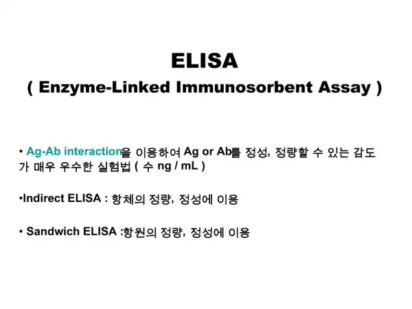
Enzyme-linked Immunosorbent Assay (ELISA): Test antigens from specimens are passed through a tube (or a membrane) coated with the corresponding specific known antibodies and become trapped on the walls of the tube (or on the membrane). Known antibodies to which an enzyme has been chemically attached are then passed through the tube (or membrane) where they combine with the trapped antigens. Substrate for the attached enzyme is then added and the amount of antigen-antibody complex formed is proportional to the amount of enzyme-substrate reaction as indicated by a color change. - Indirect ELISA - Sandwich ELISA - Competitive ELISA - Chemiluminescence
Western Blot The western blot (alternatively, proteinimmunoblot) is an analytical technique used to detect specific proteins in a given sample of tissue homogenate or extract. It uses gel electrophoresis to separate native or denatured proteins by the length of the polypeptide (denaturing conditions) or by the 3-D structure of the protein (native/ non-denaturing conditions). The proteins are then transferred to a membrane (typically nitrocellulose or PVDF), where they are probed (detected) using antibodies specific to the target protein.
Immunostaining- Immunoflourescence • -Immuno-gold EM • Immunoflourscence: A laboratory technique to identify specific antibodies or antigens. Antibody identification is usually performed on blood (serum). • Antibody tagged with flourescent dye • Antibody attached specifically to antigen • View specimen under exciting light • Flourscence microscope
Immunogold Electron Microscopy : Same principle as immunoflourscenceGold particles attached to antibodies (nanometer size particles) Viewed under EM to localise specific proteins or antigens
Flow Cytometry • is commonly used in the clinical laboratory to identify and enumerate cells bearing a particular antigen. Cells in suspension are labeled with a fluorescent tag by either direct or indirect immunofluorescence. The cells are then analyzed on the flow cytometer.In a flow cytometer, the cells exit a flow cell and are illuminated with a laser beam. The amount of laser light that is scattered off the cells as they passes through the laser can be measured, which gives information concerning the size of the cells. In addition, the laser can excite the fluorochrome on the cells and the fluorescent light emitted by the cells can be measured by one or more detectors.
Flow Cytometry FACS: Fluoresence-activated Cell sorter
Immunoelectron microscopy An electron microscope is a type of microscope that uses electrons to illuminate a specimen and create an enlarged image. Electron microscopes have much greater resolving power than light microscopes and can obtain much higher magnifications.