Download
1 / 65
650 likes | 928 Views
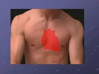
heart – surface anatomy. name these. right atrium left atrium right ventricle left ventricle. the heart. 1. Innominate (brachiocephalic) artery 2. Aortic arch 3. Ligamentum arteriosum 4. Superior vena cava 5. Right atrium 6. Right coronary artery 7. Right atrium

E N D
name these • right atrium • left atrium • right ventricle • left ventricle
the heart 1. Innominate (brachiocephalic) artery 2. Aortic arch 3. Ligamentum arteriosum 4. Superior vena cava 5. Right atrium 6. Right coronary artery 7. Right atrium 8. Inferior vena cava 9. Apex of heart 10. Right ventricle 11. Left coronary artery 12. Left ventricle 13. Left atrium 14. Pulmonary trunk 15. Left subclavian artery 16. Left common carotid artery
posterior heart 1. Left common carotid a. 2. Left subclavian a. 3. Aortic arch 4. Left pulmonary artery 5. Left pulmonary veins 6. Cut edge of pericardium 7. Left atrium 8. Inferior vena cava 9. Right atrium 10. Right pulmonary veins 11. Right pulmonary artery 12. Superior vena cava 13. Innominate (brachiocephalic) artery
heart dissection • Superior and inferior vena cava into right atrium • Rt ventricle and pulmonary artery • Pulmonary veins and Lt atrium • Lt ventricle
name the part with* Interventricular septum Interventricular sulcus Left atrioventricular valve Ligamentum arteriosum Papillary muscle Pectinate muscles Pulmonary trunk Pulmonary valve Pulmonary vein Right atrioventricular valve Septomarginal trabeculum Superior vena cava
and this * is • bicuspid valve • tricuspid valve • pulmonary semilunar valve • aortic semilunar valve
CT of heart • Right atrium • Right ventricle • Left ventricle • Left atrium • Descending aorta
impulse-conducting system 1. Sinus node (sinoatrial node) 2. Atrioventricular node 3. Right atrium 4. Right ventricle 5. Left ventricle 6. Bundle of His (atrioventricular) 6a. Right branch 6b. Left Branch 7. Left atrium
blood functions • Blood performs two major functions: transport through the body of • oxygen and carbon dioxide • food molecules (glucose, lipids, amino acids) • Ions (e.g., Na+, Ca2+, HCO3-) • wastes (e.g., urea) • hormones • heat • defense of the body against infections and other foreign materials. All the WBCs participate in these defenses.
blood is a liquid tissue Suspended in the watery plasma are seven types of cells and cell fragments. • red blood cells (RBCs) or erythrocytes • platelets or thrombocytes • five kinds of white blood cells (WBCs) or leukocytes • Three kinds of granulocytes • neutrophils • eosinophils • basophils • Two kinds of leukocytes without granules in their cytoplasm • lymphocytes • monocytes
blood • If one takes a sample of blood, treats it with an agent to prevent clotting, and spins it in a centrifuge, • the red cells settle to the bottom • the white cells settle on top of them forming the "buffy coat".
spun blood When you spin blood in a centrifuge, the red cells go to the bottom of the container, and the white cells and platelets to the middle, leaving the yellowish plasma at the top.
Plasma is the straw-colored liquid in which the blood cells are suspended. Composition of blood plasma Component- Percent • Water~92 • Proteins 6-8 • Salts 0.8 • Lipids 0.6 • Glucose (blood sugar) 0.1
plasma Plasma transports materials needed by cells and materials that must be removed from cells: • various ions (Na+, Ca2+, HCO3-, etc. • glucose and traces of other sugars • amino acids • other organic acids • cholesterol and other lipids • hormones • urea and other wastes
red blood cells (erythrocytes)the most numerous type in the blood and are responsible for the transport of oxygen and carbon dioxide • Women average about 4.8 million of these cells per cubic millimeter (mm3; which is the same as a microliter [µl]) of blood). • Men average about 5.4 x 10⁶ per µl. • These values can vary over quite a range depending on such factors as health, and altitude. (Peruvians living at 18,000 feet may have as many as 8.3 x 10⁶ RBCs per µl.)
whole blood 1. Plasma 50 % 1a. Water 90 % (45 %) 1b. Proteins 8 % (4 %) 1c. Organic acids 1 % (0.5 %) 1d. Salts 1 % (0.5 %) 2. Blood cells 45 % 2a. Erythrocytes 44 % 2b. Leukocytes & platelets 1 %
white blood cells • are much less numerous than red (the ratio between the two is around 1:700), • have nuclei, • participate in protecting the body from infection, • consist of lymphocytes and monocytes with relatively clear cytoplasm, and three types of granulocytes, whose cytoplasm is filled with granules.
Lymphocytes After neutrophils, lymphocytes are the most numerous of the circulating leukocytes. The normal range count is 1000 - 4800/µL. Their life span may vary from several days to a lifetime (as for memory lymphocytes). Lymphocytes 1) can move back and forth between the vessels and the extravascular tissues, 2) are capable of reverting to blast-like cells, and 3) when so transformed, can multiply as the immunologic need arises.
lymphocytes Each of the white blood cell types has a task in helping the body fight infections. The lymphocytes help create antibodies that attack the invaders and mark them for destruction by the neutrophils, monocytes and macrophages.
monocytes largest cell type seen in blood smears, and constitute 5 to 8% of total leukocytes. Their nuclei are not multilobular like granulocytes, but may be deeply indented or U-shaped
monocytes The normal range for the monocyte count is 200 - 950 /µL.
neutrophils- the most abundant WBC Neutrophils squeeze through the capillary walls and into infected tissue where they kill the invaders (e.g., bacteria) and then engulf the remnants by phagocytosis.
eosinophils The number of eosinophils in the blood is normally quite low (0 - 450/µl). However, their numbers increase sharply in certain diseases, especially infections by parasitic worms. Eosinophils are cytotoxic, releasing the contents of their granules on the invader.
basophils • The number of basophils also increases during infection. Basophils leave the blood and accumulate at the site of infection or other inflammation. There they discharge the contents of their granules, releasing a variety of mediators such as: histamine, serotonin prostaglandins, and leukotrienes
basophils increase blood flow to the area and in other ways add to the inflammatory process. The mediators released by basophils also play an important part in some allergic responses such as hay fever and to insect stings.
5. Leukocytes (white blood corpuscles) 5a. Neutrophil (granulocyte) 5b. Eosinophil (granulocytes) 5c. Basophil (granulocyte) 5d. Monocyte 5e. Small lymphocyte 5f. Large lymphocyte blood cells 3. Erythrocytes (red blood cells) 4. Thrombocytes (platelets) 5. Leukocytes (white blood cells) 5a. Neutrophil (granulocyte) 5b. Eosinophil (granulocytes) 5c. Basophil (granulocyte) 5d. Monocyte 5e. Small lymphocyte 5f. Large lymphocyte
platelets Blood normally contains 150,000 to 450,000 per microliter (µl). If this value should drop much below 50,000/µl, there is a danger of uncontrolled bleeding. This is because of the essential role that platelets have in blood clotting. Look for the t’s, platelet beside each.
Blood Group Blood Group Antigens on RBCs Antigens on RBCs Antibodies in Serum Antibodies in Serum Genotypes Genotypes A A A A Anti-B Anti-B AA or AO AA or AO B B B B Anti-A Anti-A BB or BO BB or BO AB AB A and B A and B Neither Neither AB AB O O Neither Neither Anti-A and anti-B Anti-A and anti-B OO OO ABO’s of blood The table shows the four ABO phenotypes ("blood groups") present in the human population and the genotypes that give rise to them.
wrong transfused blood Human red blood cells before (left) and after (right) adding serum containing anti-A antibodies. The agglutination reaction reveals the presence of the A antigen on the surface of the cells.
Rh factor This protein is also present in the blood of some people. Other people, however, do not have the protein. The presence of the protein, or lack of it, is referred to as the Rh (for Rhesus) factor. If your blood does contain the protein, your blood is said to be Rh positive (Rh+). If your blood does not contain the protein, your blood is said to be Rh negative (Rh-).
blood type and Rh factor This Rh factor is connected to your blood type. For example, your blood may be AB+ which means that you have type AB blood with a positive Rh factor. Or, you might have O- blood which means that you have type O blood with a negative Rh factor.
mother and fetus’ Rh factor It is particularly important for expectant mothers to know their blood's Rh factor. Occasionally, a baby will inherit an Rh positive blood type from its father while the mother has an Rh negative blood type. The baby's life could be in great danger if the mother's Rh negative blood attacks the baby's Rh positive blood. If this happens, an exchange transfusion may save the baby's life. The baby's blood can be exchanged for new blood that matches the mother's.
blood vessels • arteries • arterioles • capillaries • venules • veins
diagram of capillary network 1. Arteries 2. Arterioles 3. Capillaries 4. Venules 5. Veins
arteries • heart pumps blood out through one main artery called the dorsal aorta which divides and branches out into many smaller arteries so that each region of your body has its own system of arteries supplying it with fresh, oxygen-rich blood • arteries are tough on the outside and smooth on the inside, have actually three layers: an outer layer of tissue, a muscular middle, and an inner layer of epithelial cells
artery and vein 1. Lumen 2. Tunica initima 3. Endothelium of tunica initima 4. Internal elastic membrane 5. Tunica media 6. Smooth muscle cells of tunica media 7. External elastic membrane 8. Tunica adventitia 9. Longituduinal cells of adventitia 10. Fibre lattice of adventitia 11. Vasa vasorum 12. Valves
capillaries • are very thin and fragile. The capillaries are actually only one epithelial cell thick • so thin that blood cells can only pass through them in single file • exchange of oxygen and carbon dioxide takes place through the thin capillary wall • red blood cells inside the capillary release their oxygen which passes through the wall and into the surrounding tissue • tissue releases its waste products, like carbon dioxide, which passes through the wall and into the red blood cells
veins • veins are similar to arteries but, because they transport blood at a lower pressure, they are not as strong as arteries • like arteries, veins have three layers: an outer layer of tissue, muscle in the middle, and a smooth inner layer of epithelial cells • the layers are thinner, containing less tissue. Veins receive blood from the capillaries after the exchange of oxygen and carbon dioxide has taken place • veins transport waste-rich blood back to the lungs and heart. • valves located inside the veins prevent the “backflow” of blood
circulation 1. Heart and arms 2. Superior vena cava 3. Lungs 4. Right atrium 5. Right ventricle 6. Liver 7. Portal vein 8. Inferior vena cava 9. Kidneys 10. Legs 11. Intestines 12. Abdominal aorta 13. Left ventricle 14. Left atrium 15. Pulmonary veins 16. Pulmonary artery
path of circulation 1. 2 & 10 sup/inf vena cava 2. 7 Rt atrium 3. 8 Rt AV valve (tricuspid) 4. 9 Rt ventricle 5. 6 Rt SL (semilunar) valve 6. 4,3,18 Pulmonary arteries 7. lungs 8. 5, 17 Pulmonary veins 9. 16 Lt atrium 10. 14 Lt AV valve (bicuspid) 11. 13 Lt ventricle 12. 15 Lt SL (semilunar) valve 13. 1 aorta
dub Diastole (relaxation of heart muscle) bottom number of B.P. Such as 120/80 lub Systole (contraction of the heart muscle) top number of B.P. Such as 120/80 blood pressure heart beats…lub dub
heart valves from above 1. Tricuspid (right atrioventricular) valve 2. Aortic valve (Lt SL) 3. Pulmonary valve (Lt SL) 4. Mitral (bicuspid or left atrioventricular) valve
pulmonary circulation pulmonary circulation is the movement of blood from the heart, to the lungs, and back to the heart again