Download
1 / 30
320 likes | 831 Views
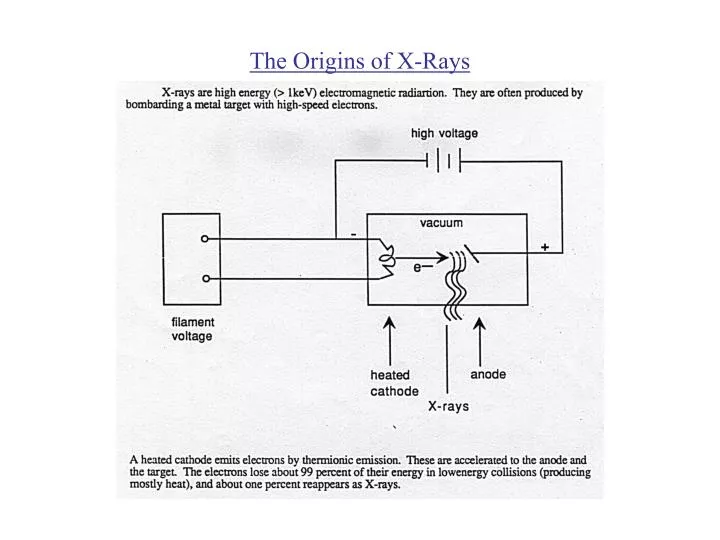
The Origins of X-Rays. The X-Ray Spectrum. The X-Ray Spectrum (Changes in Voltage). The continuous spectrum is from electrons decelerating rapidly in the target and transferring their energy to single photons, Bremsstrahlung.

E N D
The X-Ray Spectrum (Changes in Voltage) The continuous spectrum is from electrons decelerating rapidly in the target and transferring their energy to single photons, Bremsstrahlung. The characteristic lines are a result of electrons ejecting orbital electrons from the innermost shells. When electrons from outer shells fall down to the level of the inner ejected electron, they emit a photon with an energy that is characteristic to the atomic transition.
The X-Ray Spectrum (Changes in Target Material) Increase in Z: Increase in X-ray intensity since greater mass and positive charge of the target nuclei increase the probability of X-ray emission total output intensity of Z Characteristic lines shift to higher energy, K and L electrons are more strongly held No change in
The X-Ray Spectrum Filtrations typically one wishes to remove low-energy X-rays from the beam. This is accomplished by placing a sheet of metal in the path of the X-ray beam. Changes the X-ray spectrum shape by removing low-energy electrons Shifts the spectrum peak to higher energies Reduces the overall X-ray output Shifts Emin to higher energies No change in Emax.
Beam Hardening The beam from an X-ray source is not mono-energetic and the lower energy photons will be more attenuated than the higher energy ones.
Image of Focal Spot Using A Pinhole Scan picture
Source Considerations In X-ray Imaging Cathode is finite in size.
Source Considerations In X-ray Imaging Notice that as is reduced the loading efficiency increases, but the angular width of the beam decreases. Typical spot size for planar imaging
Source Considerations In X-ray Imaging Width = 16, the effective spot size is reduced to
Source Considerations In X-ray Imaging Heel Effect Intensity of Beam with Angle
Source Considerations In X-ray Imaging The true spot on an anode is inside the anode. Why not use larger angles? Greater spot size. What about X-ray spectrum vs. angle?
Source Considerations In X-ray Imaging Schematic of calculation
Scatter Analysis #1 The incremental density of the scattered photons generated in the plane at height z is:
Scatter Analysis #2 • It is not enough to know the number of photons scattered, we also need to know how many are scattered towards the detector. • at diagnostic energy ???, the fraction forward scattered, k • the number that reaches the detector is • if only 1 scatter event per photon
Scatter Analysis #4 But this is not the entire picture, we know that there are multiple scatter events for individual photons. The mean distance traveled along z for forward directed particles before a scatter event is: The average number of interactions along a length L is:
Scatter Analysis #5 The ratio of scattered to transmitted photons is:
Poisson Density Function As we have seen, X-rays are discrete photons. The probability that exactly k photons will be emitted over a definite period in time is given by the Poisson density function. A defining feature of the Poisson distribution is that the variance, 2, (or the central second moment - width) is equal to the mean.
Poisson Density Function The signal-to-noise of a measurement X-ray photons is then: signal o where = average # of photons Noise root mean square deviation from kEo Consider the effect of an energy spectrum for the S/N. Detection efficiency generally goes as the stopping power, therefore lower for higher energy photons .
Types of Noise (Additive Noise) Additive Noise - When the energy photons is low then there are many photons and they may be thought of as arriving continuously. There are virtually no statistical fluctuations in the arrival rate, only Johnson type noise added by the measurement system.
Types of Noise (Quantum Noise) Quantum noise (“counting” noise) - high energy per photon, therefore only a few photons are required but now since each photon can be detected individually and the counting rate is low, there are statistics associated with the arrival of the photon at the detector.
Photon Statistics So for X-rays So S/N depends on the counting statistics of photons reading the detector. Outline of proof that photons energy from a material continue to follow Poisson statistics. The emission of X-ray from a source follow Poisson statistics.
Photon Statistics Interactions of photons with matter is a binary process. They interact or not (ideal case), therefore it is a binomial process. Put these two together to find the probability of sending k photons through an object, Q(k). probability of photon source generating k+n photons probability of n photons being transmitted # of permutations of sucn an event probability of k photons being transmitted
Poisson Distribution Photons emerging from an attenuating object continue to follow a Poisson distribution, however with the rate scaled by the attenuation. Note: True for an all or nothing process. The photons emitted have a mean value. Clearly S/N is increased at the cost of dose.