Download
1 / 8
270 likes | 1.06k Views
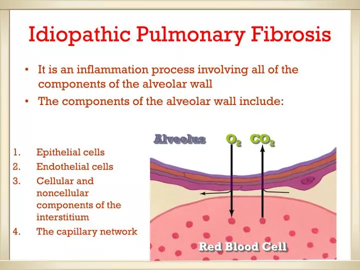
Idiopathic Pulmonary Fibrosis. It is an inflammation process involving all of the components of the alveolar wall The components of the alveolar wall include:. Alveolus. O 2. CO 2. Epithelial cells Endothelial cells Cellular and noncellular components of the interstitium

E N D
Idiopathic Pulmonary Fibrosis • It is an inflammation process involving all of the components of the alveolar wall • The components of the alveolar wall include: Alveolus O2 CO2 • Epithelial cells • Endothelial cells • Cellular and noncellular components of the interstitium • The capillary network Red Blood Cell
Pathophysiology Inadequate O2 O2 • Reduced lung compliance increase work of breathing (WOB) • V/Q mismatching • Impaired diffusion of oxygen to alveolar capillary • Hypoxemia • Respiratory failure O2 Wasted perfusion
Clinical Manifestation • Insidious or acute onset dyspnea on exertion (DOE) progress to resting dyspnea • Repetitive nonproductive cough • Fatigue • Loss of appetite, weight loss • Tachypnea
Clinical Manifestation • Diffuse reticulonodular pattern in the involved areas on chest x-ray (patient may have a normal x-ray) • Decreased PO2, during exercise, later at rest, and normal PCO2 • Normal PO2= 80-100, Normal PCO2= 35-45
Clinical Manifestation • An abnormal chest x-ray shows scarring and cyst formation in both lungs, predominantly in the middle and lower areas. These findings are typical of idiopathic pulmonary fibrosis. A normal chest x-ray is shown on the right for comparison; the heart (H), lungs (L), vertebrae (v), and clavicle (C) can be seen.
Clinical Manifestation • Bibasilar end-inspiratory rales, and decreased breath sounds Click here to listen to an IPF patient’s breath sound
Clinical Manifestation • Cyanosis later • Digital clubbing • Cor-polmonale or respiratory failure
Treatment • Corticosteroids • Cytoxic drugs e.g. cyclophosphamide and azathioprine • Combine 1&2 treatment is preferable • Supportive measures e.g. smoking cessation, oxygenation and ventilation, good nutrition, and aggressive treatment of infection