Download
1 / 39
410 likes | 571 Views
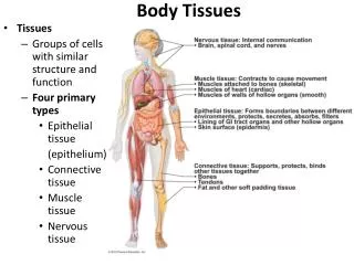
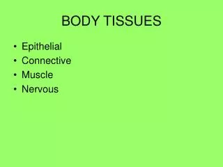
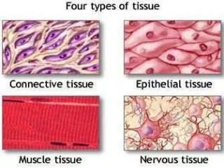
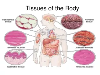
Body Tissues rev 9-11 Tissue : group of cells that are similar in structure and perform a common function 4 primary tissue groups Epithelial Connective Muscle Nervous Epithelial Tissue sheet of cells that covers a body surface or lines a body cavity;

E N D
Body Tissues rev 9-11 • Tissue: group of cells that are similar in structure and perform a common function • 4 primary tissue groups • Epithelial • Connective • Muscle • Nervous Tissues & Skin
Epithelial Tissue • sheet of cells that covers a body surface or lines a body cavity; • helps form boundaries between different body environments • protects underlying tissues • reduces friction because it is smooth • absorbs • secretes Tissues & Skin
Forms glands • Characteristics of Epithelial Tissue 1. Cells are packed closely together 2. One surface of the tissue is free while the other surface is attached to the underlying connective Tissues & Skin
3. Epithelial tissues are typically given 2 names--first name indicates the number of cell layers present • one layer of cells is called simple epithelium • many layers--stratified epithelium • second name describes the shape of the cells Tissues & Skin
4. Cells occur in 3 types • squamous--thin and flat cells • cuboidal--cube or rounded cells • columnar--cylindrical cells • these tissues may also have microvilli, cilia, or goblet cells Tissues & Skin
Basement Membrane • Underneath the cells of epithelial tissue is a supporting non-cellular layer called the basement membrane and beneath that is typically a layer of connective tissue. • Epithelial cells can also be attached to each other by different types of cell junctions: • Tight junctions, • Adhesive junctions, • Gap junctions Tissues & Skin
Tight junctions: seal plasma membranes of adjacent cells so tightly that nothing can pass between the cells • Adhesion junctions: (also called spot desmosomes) are looser in structure and allow for some movement between cells so the tissues can stretch and bend • Gap junctions: are connecting protein channels that permit the movement of ions or water between 2 adjacent cells Tissues & Skin
Connective Tissue found everywhere in the body Major functions: 1. Binding or connecting of body parts 2. Support of organs against gravity 3. Protection 4. Cushioning, insulation; energy storage; fat storage 5. Produces blood cells 6. Transportation • Has comparatively few cells and a lot of matrix Tissues & Skin
Types of Connective tissue: • Fibrous-connects various body parts; provides strength, support and flexibility • consists of several types of fibers and cells embedded in a gel-like ground substance: • Collagen fibers--provides strength and slight flexibility • Elastic—thin and very flexible coiled elastic fibers made from the protein elastin • Reticular—made of thinner collagen fibers which interconnect with each other; serves as internal framework for some organs; fiber flexibility is between elastic and collagen fibers Tissues & Skin
The various fibers are set in a ground substance (called the matrix) • It contains many types of cells including fat cells, mast cells, various WBC, and fibroblasts. Tissues & Skin
Fibrous connective tissues are subclassified according to the density and arrangement of their fibers: Loose Areolar connective tissue • most common type • contains collagen fibers and elastic fibers in a “loose” irregular pattern; is very flexible but not strong • usually found below the skin, between muscles, and around blood vessels, muscles and organs. • Spaces between fibers are good storage areas. Tissues & Skin
Dense (Regular) Connective Tissue • fibers are densely packed and run in the same direction • is very strong when stress is in the same direction as the fibers run • has few blood vessels and takes a long time to heal • functions to bind, protect and connect Tissues & Skin
Elastic Connective Tissue: • Surrounds organs that have to reguarly change shape or size • Contains a high proportion of elastic fibers Reticular connective Tissue: • Also called lymphoid tissue • Serves as the internal framework of soft organs such as the liver, lymphatic system organs • Is made up of thin, branched reticular fibers Tissues & Skin
Specialized connective tissues: Cartilage: is the transition tissue from which bone develops • Produced by chondroblasts which become trapped and enclosed in areas called lacunae; no blood vessels, high collagen fiber and water content • Because there are no blood vessels, mature cells obtain nutrients by diffusion through the ground substance. • Maintains the shape of certain body parts • Protects and cushions joints; cartilagenous disks cushion the vertebrae, forms the tough covering of bones at joints Tissues & Skin
Bone: connective tissue which contains only a few living cells • inorganic matrix with calcium and phosphate salts for hardness Blood: cells are suspended in a fluid matrix called plasma. Considered a connective tissue because all blood cells derive from earlier stem cells located within bone. • Red blood cells transport oxygen and nutrients • White blood cells (WBC) function in the immune system • Platelets help to form blood clots following an injury. Tissues & Skin
Adipose (Fat) tissue: specialized for fat storage • has few connective tissue fibers and almost no ground substance; primarily made up of adipocytes (fat cells) • Primary role is insulation and cushioning; stores energy; forms a protective layer around internal organs Tissues & Skin
Muscle Tissue: Contracts for Movement Muscle tissue is made up of tightly packed cells called muscle fibers. The muscle fiber cytoplasm contains proteins which allow the cell to contract 3 types of muscle tissue • Skeletal muscle moves body parts. • Is connected to tendons which are connected to bones. • Voluntary, multinucleated Tissues & Skin
Cardiac muscle: • Found only in the heart • Individual cells are shorter than skeletal, have single nucleus • Cells are arranged parallel to each other • Have intercalated disks which function as gap junctions for direct electrical contact with neighboring cells. This allows one cell to activate all its “neighbors” so the heart can contract in a coordinated way. • Involuntary muscle Tissues & Skin
Smooth muscle: • Surrounds hollow organs and tubes i.e. blood vessels, digestive tract • Smaller cells than skeletal muscles; have a single nucleus • Cells arranged parallel to each other • Have gap junctions between cells so that it works in a coordinated fashion • Involuntary muscle Tissues & Skin
Nervous Tissue: Transmit Impulses • Nervous tissue is made up of cells which are specialized for generating and transmitting electrical impulses. It is a rapid communication network for the body. • Neuron: specialized nervous system cell which generates and transmits impulses. • Structural components: cell body, dendrites, axon • Glial cells: support neuron cells and supplies with nutrients Tissues & Skin
INTEGUMENTARY SYSTEM (SKIN) Functions: --outer covering of the body • protects from dehydration • protects from injury • protects against invasion by microorganisms (bacteria and viruses • helps regulate body temperature • synthesizes vitamin D • Sensory awareness: receptors for touch, vibration, pain and temperature provide information about the environment Tissues & Skin
Skin consists of • epidermis: outermost layer of stratified squamous epithelial tissue • is made up of 5 sub-layers: stratum corneum, stratum lucidum, stratum granulosum, stratum spinosum, and stratum basale layers Tissues & Skin
Innermost layer: stratum basale undergoes almost continuous mitosis. Cells are pushed upward by the production of new cells beneath them and create “new” skin • Melanocytes are found here; they produce melanin, a brown pigment. • Stratum spinosum: thickest layer; cells switch from a mitotic role to producing keratin. • Keratin is a waterproofing protein which also toughens the outer surface of the skin. • Macrophages (phagocytes which protect us from infection) are also present throughout this skin layer Tissues & Skin
Stratum granulosum: acts as a protective shield to the layers below it. • Stratum lucidum: thin layer with keratin production occurring • Stratum corneum: outermost and toughest layer of epidermis • Outermost layers of epidermis are made up of dead, dried out epithelial cells which contain keratin • When cells are dead and water has evaporated, keratin forms a tough barrier • Provides protection from abrasion, cells can be rubbed off and will be replaced; also protects our body from drying out Tissues & Skin
The dermis is primarily dense connective tissue with collagen, elastic and reticular fibers in a ground matrix. • The fibers allow the skin to stretch when we move • give it strength to resist abrasion and tearing. • Our skin becomes less flexible and more wrinkled as we age. Tissues & Skin
composed of two sub-layers • this layer “binds” the body together • richly supplied with nerve fibers, blood vessels, hair follicles, sebacious (oil) glands, sweat glands and lymphatic vessels • Sensory nerve endings: for heat, cold, touch, deep pressure, vibration; provide information about the outside environment • Nerve fibers:Meissner’s corpuscles-light touch Pacinian corpuscles-deep pressure Free nerve endings-pain Tissues & Skin
There are 2 types of sweat glands: • Eccrine which are throughout the body and • Apocrine which are primarily in the groin and underarm (axillary) areas. Sweat helps in temperature regulation and contains an antibiotic called dermicidin. • Arrector pili muscles which make our hair stand up • Blood vessels: supply nutrients, remove waste, assist in temperature regulation • Nails: a scale like modification of the epidermis • Ceruminous or wax gland Tissues & Skin
Hypodermis: supportive layer consisting of loose connective tissue containing fat cells • also called subcutaneous tissue or superficial fascia • anchors the skin to underlying structures • is flexible so the skin can move and bend • its fat cells insulate against excessive heat loss and cushion against injury Tissues & Skin
Diseases of the Skin Impetigo • A contagious, superficial infection in bullous (blister like) and nonbullous forms • Usually occurs on face, around the mouth and nose • ITCHY! • Causes: • Staph aureus usually causative organism • When blister breaks, liquid (exudate) can cause more lesions on rest of body Tissues & Skin
Treatment: • Antibiotics (penicillin, cephalosporin, zithromax • Anti-itch cream—itching spreads impetigo • Frequent washing of lesions with antibacterial soap • Patient has own towels, bedding, etc • Caretaker must wash hands carefully and frequently Tinea (Skin Fungus) infections: • can occur directly (through contact with infected lesions) or indirectly (through contact with contaminated articles-shoes, towels, or shower stalls Tissues & Skin
Tinea capitis • Small, spreading blister like rash on scalp causing patchy hair loss and scaling • Usually affects children; in babies called “cradle cap” • Tinea corporis (also known as ringworm): • produces flat lesions on the skin which, as they get bigger, have healed centers and look like a ring • Tinea pedis (Athlete’s foot) • Scaling and blisters between the toes Tissues & Skin
Tinea cruris (Jock itch) • Produces red, raised, sharply defined, itchy lesions in the groin that can extend to the buttocks, inner thighs, and the external genitalia. • Warm weather and tight clothing encourage fungal growth • Treatment for all Tineas • Usually topical creams • Continue applying cream for 2 weeks after lesions heal • Observe for secondary infections • Expose areas (when possible) to air Tissues & Skin
Scabies • Infection by the “itch mite” which causes a sensitivity reaction • Occurs primarily in areas with overcrowding and poor hygiene • Very contagious; transmitted through skin or sexual contact • Mite lives in the skin. Female burrows into the skin to lay her eggs. The larvae emerge to copulate and then reburrow under the skin Tissues & Skin
Causes itching which intensifies at night and can lead to a secondary bacterial infection • Lesions are usually excoriated, threadlike, about 3/8 inch long and typically seen between fingers, on flexor surfaces of wrist, on elbows, underarm, at the waistline, and can be seen in genitalia Treatment: • Cream over entire skin surface and left on for 8-12 hours to 5 days (depending on the specific type of cream) • Application usually repeated in 1 week • Oral antihistamine • Wash clothes and bedding in very hot water or dryclean Tissues & Skin
Lice or Pediculosis • Pediculus capitis (head lice) • Pediculus corporis (body lice) • Pediculus pubis (crab lice) • Lice feed on human blood and lay their eggs (nits) in body hairs or clothing fibers • After nits hatch, lice must feed within 24 hours or die. Mature in 2-3 weeks • When louse bites it injects a toxin into the skin that produces irritation and a purpuric spot (spot containing leaked blood). Tissues & Skin
Causes itching which can cause skin breakdown, swollen lymph glands, rash • Treatment: • Special shampoo, creams • Shampoo applied and washed off after 5-10 minutes (depending on specific shampoo); repeat in 7-10 days • After this, all nits should be combed out of hair with a metal nit comb • Wash clothing and bedding Tissues & Skin
Psorias • Chronic disease marked by epidermal proliferation; skin is covered by scales • Life cycle of normal skin is ~ 28 days and then if sloughs off; life cycle of psoriatic skin is 4 days so the cell can’t mature resulting in thick, flaky skin • Has remissions and exacerbations • Tendency to develop this is genetic • Symptoms: itching; dry, cracked skin lesions which can cause pain. • Lesions commonly appear on scalp, chest, elbows, knees, shins, back, and buttocks Tissues & Skin
Treatment: • No permanent cure exists • Lotions or tar preparations to help soften the scales which then can be scrubbed off gently; ultraviolet light to retard rapid cell production; • Steroid creams to control disease • Antihistamines to relieve itching • Not contagious Tissues & Skin
Eczema • Chronic inflammatory response often associated with allergies, irritating chemicals, temperature and humidity, skin irritation (i.e. tight clothing), and emotions. • Lesions generally begin as raised areas on skin • Typically flares and subsides repeatedly • During flareups, itching and scratching can cause edema, crusting and scaling • Treatment: eliminate allergens and irritations • Antihistamines to relieve itching, steroid creams; Hydrate skin; Short baths/showers in cool water Tissues & Skin