Download
1 / 81
1.43k likes | 4.31k Views
Renal Failure. Renal Failure. Results when kidney’s cannot remove wastes or perform regulatory functions Systemic disorder resulting from many different causes ARF : Reversible syndrome that results in decreased GFR and oliguria (Fast & Days )

E N D
Renal Failure • Results when kidney’s cannot remove wastes or perform regulatory functions • Systemic disorder resulting from many different causes • ARF : Reversible syndrome that results in decreased GFR and oliguria (Fast & Days ) • CRF : Progressive ( Slow & Years ); Irreversible deterioration of renal function resulting in azotemia • Both types result in loss of ability to maintain normal volume and composition of body fluids
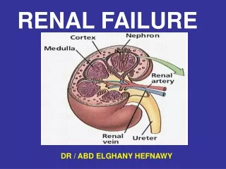
Assessment of Renal Function • Glomerular Filtration Rate (GFR) • = The volume of water filtered from the plasma per unit of time. • Gives a rough measure of the number of functioning nephrons • Normal GFR: • Men: 130 mL/min./1.73m2 • Women: 120 mL/min./1.73m2 • Cannot be measured directly, so we use creatinine and creatinine clearance to estimate.
Assessment of Renal Function • Creatinine • As plasma creatinine increases, the GFR exponentially decreases. • Limitations to estimate GFR: • Patients with decrease in muscle mass,, malnutrition, advanced age, may have low/normal creatinine despite underlying kidney disease • Medications may artificially elevate creatinine: • Trimethroprim • Cimetidine
Assessment of Renal Function • Creatinine Clearance • Best way to estimate GFR • GFR = (creatinine clearance) x (body surface area in m2/1.73) • Ways to measure: • 24-hour urine creatinine: • Creatinine clearance = (Ucr x Uvol)/ plasma Cr • Cockcroft-Gault Equation: (140 - age) x lean body weight [kg] CrCl (mL/min) = ——————————————— x 0.85 if Cr [mg/dL] x 72 female
Major causes of Kidney Failure • Prerenal Disease • Vascular Disease • Glomerular Disease • Interstitial/Tubular Disease • Obstructive Uropathy
Acute Renal Failure Relatively sudden onset of renal failure that is potentially reversible
Acute Renal Failure • An abrupt decrease in renal function sufficient to cause retention of metabolic waste such as urea and creatinine. • Most community acquired acute renal failure (70%) is prerenal • Most hospital acquired acute renal failure (60%) is due to ischemia or nephrotoxic tubular epithelial injury (acute tubular necrosis). • Mortality rate 50-70% • Risk factor for acute renal failure • Advanced age • Preexisting renal parenchymal disease • Diabetes mellitus • Underlying cardiac or liver disease • Early sign of ARF is oliguria. • Only seen in 2/3 of ARF pts. • Frequently have: • Metabolic acidosis • Hyperkalemia • Disturbance in body fluid homeostasis • Secondary effects on other organ systems
Definition of Acute Renal Failure Based on “Acute Kidney Injury Network”
RIFLE criteria for diagnosis of ARF based on The “Acute Dialysis Quality Initiative”
Classification of ARF Acute Renal Failure Pre-renal Intrinsic Post-renal Glomerular Interstitial Tubular Vascular
Acute Renal Failure Pre Renal Causes Intrinsic causes Post Renal Causes Tubular Interstitial Acute Necrosis Nephritis Glomerulonephritis (10% of cases) (5% of cases) Ischemia Toxins (50% of cases) (35% of cases)
Ischemic Acute Renal Failure Intravascular volume depletion and hypotension Gastrointestinal, renal, dermal losses, hemorrhage, shock Decreased effective intravascular volume:CHF, cirrhosis, nephrosis, peritonitis Large vessel renal vascular disease: Renal artery thrombosis or embolism, Renal artery stenosis Generalized or localized reduction in renal blood flow Medications:ACE inhibitors, NSAIDS, radiocontrast agents, Ampho B, Cyclosporin Small vessel renal vascular disease: Atheroembolism, vasculitis, malignant hypertension, hypercalcemia, transplant rejection Hepatorenal syndrome Sepsis Ischemic Acute Renal Failure
Effects of NSAIDS and ACE inhibitorson Glomerular Hydrostatic Pressure Prostaglandins cause afferent vaso dilation Locally produced AII causes efferent vaso constriction Blocked by ACE inhibitors Blocked by NSAIDS
Most common causes of ACUTE Renal Failure • Prerenal ( Community ) • Acute Tubular Necrosis (ATN) – ( Hospitals ) • Acute on chronic renal failure (usually due to ATN or prerenal) • Obstructive uropathy • Glomerulonephritis/Vasculitis • Acute Interstitial nephritis • Atheroemboli
Prerenal Azotemia Intravascular volume depletion Bleeding, GI loss, Renal loss, Skin loss, Third space loss Decreased cardiac output CHF Renal vasoconstriction Liver Disease, Sepsis, Hypercalcemia Pharmacologic impairment of Autoregulation and GFR in specific settings ACEI in bilateral RAS, NSAIDS in any renal hypoperfusion setting
Acute Tubular Necrosis(ischaemic or toxic) [ATN] Interstitial nephritis Glomerular disease Renal Vascular Disease Microvascular Vasoconstriction Tubular Obstruction 70% ATN Drugs (Aminoglycosides,NSAIDs, Amphotericin) Rhabdomyolysis Iodinated contrast agents Heavy metals Prolonged ischaemia Combination of the above Renal Acute Renal Failure
Intrinsic Renal Azotemia Large Renal Vessel Disease Thrombo-embolic disease Renal Microvasculature and Glomerular Disease Inflammatory: glomerulonephritis, allograft rejection Vasospastic:malignant hypertension, scleroderma crisis, pre-eclampsia, contrast Hematologic: HUS-TTP, DIC Acute Tubular Necrosis (ATN) Ischemic Toxic Tubulo-interestitial Disease Acute Interestitial Nephritis (AIN), Acute cellular allograft rejection, viral (HIV, BK virus), infiltration (sarcoid) Intratubular Obstruction myoglobin, hemoglobin, myeloma light chains, uric acid, tumor lysis, drugs (indinavir, acyclovir, foscarnet, oxalate in ethylene glycol toxicity)
Acute Tubular Necrosis • Most common form of “Renal” ARF • Tubular damage, loss of tubular function with direct effect on GFR • U/P osmol =1, U/P creatinine 10-20, UNa+ > 40 mmol/l • Duration days to 6 weeks • Oliguria most common but anuria and polyuria possible • Diuretics do not change course of ATN but can increase water excretion • High mortality even with dialysis (ATN associated with other organ failure
Postrenal Azotemia Stones Blood clots Papillary necrotic tissue Urethral disease Anatomic: posterior valve Functional: anticholinergics, L-DOPA Prostate disease Bladder disease Anatomic: cancer, schistosomiasis Functional: neurogenic bladder
Urine Output in Acute Renal failure • Oliguria • = Daily urine output < 400 mL • When present in acute renal failure, associated with a mortality rate of 75% (versus 25% mortality rate in non-oliguric patients) • Most deaths are associated with the underlying disease process and infectious complications • Anuria • No urine production • Probably time for dialysis
Phases of Acute Renal Failure • Phases of rapid decrease in renal function lead to the collection of metabolic wastes in the body. • Phases include: • Oliguric • Diuretic • Recovery • Acute syndrome may be reversible with prompt intervention.
Oliguric Phase • Clinical picture dominated by surgical, medical or obstetric problem causing ARF • Oliguria within 24-48 hours of initial injury • May take several days to develop with nephrotoxic chemicals • Azotemia accompanies oliguria • Critical to recognize, determine cause and begin treatment • Oliguria caused by acute-on-chronic RF usually easy to detect from history • Post renal obstruction must be ruled out • Prerenal oliguria most common condition leading to ARF and must be distinguished from ATN
Diuretic Phase • Begins when urine output increases to >400 ml/day • Usually lasts 2-3 weeks • Urine output rarely exceeds 4 L/day • Caused partly by osmotic diuresis due to high blood urea and partly by impaired ability of recovering tubules to conserve salts and water • May develop K+, Na+ and water deficits • Must replace loses or death • BUN may continue to rise as clearance does not keep up with production • With continued diuresis azotemia gradually disappears and get clinical improvement
Recovery Phase • Lasts up to one year • Anemia and concentrating ability gradually improve • Some have permanent reduction in GFR • ATN serious condition (still 50% mortality – down from 90% 30 years ago) • About 2/3 die during oliguric stage and about 1/3 during diuretic stage • Mortality related to cause • 60% after surgery, crushing injuries • 25% after CCl4, bad transfusion • 10-15% in obstetric cases • With non-oliguric ARF – 25% mortality
Natural Clinical Course of ATN Initiation Phase (hours to days) Continuous ischemic or toxic insult Evolving renal injury ATN is potentially preventable at this time Maintenance Phase (typically 1-2 wks) May be prolonged to 1-12 months Established renal injury GFR < 10 cc/min, The lowest UOP Recovery Phase Gradual increase in UOP toward post-ATN diuresis Gradual fall in SCr (may lag behind the onset of diuresis by several days)
ARF: Systemic Complications • Infections of urinary tract & lungs due to uremia • Up to 70% of pts. with ARF. • #1 cause of ARF morbidity/mortality • Anemia • Kidney makes EPO, ↓ EPO anemia (HCT 20-30) • “3rd space disease” • Salt and Water retention (esp. in prerenal failure) • Pulmonary edema, Pleural effusion, & ascites • Hypocalcemia • ↓ Excretion of phosphate impaired GI absorption of Calcium. • Hyperkalemia • ↓ Glomerular filtration, ↓ Tubular secretion • Malaise, nausea, and muscle weakness. • A cardiac emergency • Metabolic Acidosis w/ ↑ Anion Gap • ↓ Excretion of acids & ↓ tubular reabsorption of bicarbonate results in metabolic acidosis with a high anion gap. • Hypotension, Kussmaul’s respirations
Initial Diagnostic Tools in ARF History and Physical examination Detailed review of the chart, drugs administered, procedures done, hemodynamics during the procedures. Urinalysis (SG, PH, protein, blood, crystals, infection ) Urine microscopy {casts, cells (eosinophils)} Urine Electrolytes Renal imaging (US, CT Scan ,Retrograde Pyelogram ) Markers of CKD (iPTH, size<9cm, anemia, high phosphate, low bicarbbonate Renal biopsy
Assessing the patient with ARF • History: • Cancer? • Recent Infections? • Blood in urine? • Change in urine output? • Flank Pain? • Recent bleeding? • Dehydration? Diarrhea? Nausea? Vomiting? • Blurred vision? Elevated BP at home? Elevated sugars?
Assessing the patient with ARF • Family History: • Cancers? • Polycystic kidney disease? • Medications: • Any non-compliance with diabetic or hypertensive meds? • Any recent antibiotic use? • Any NSAID use?
Assessing the patient with ARF Physical Exam • Vital Signs: • Elevated BP: Concern for malignant hypertension • Low BP: Concern for hypotension/hypoperfusion (acute tubular necrosis) • Neuro: • Confusion: hypercalcemia, uremia, malignant hypertension, infection, malignancy • HEENT: • Dry mucus membranes: Concern for dehydration (pre-renal) • Abdomen: • Ascites: Concern for liver disease (hepatorenal syndrome), or nephrotic syndrome • Extremities: • Edema: Concern for nephrotic syndrome • Skin: • Tight skin, sclerodactyly – Sclerodermal renal crisis • Malar rash - Lupus
Assessing the patient with ARFLaboratory Analysis • Fractional Excretion of Sodium: (UrineNa+ x PlasmaCreatinine) FENa= ______________________ x 100 (PlasmaNa+ x UrineCreatinine) • FENa < 1% → Prerenal • FENa > 2% → (Acute Tubular Necrosis),obstructive uropathy • If patient receiving diuretics, can check FE of urea.
Assessing the patient with ARF Radiology • Renal Ultrasound • Look for signs of hydronephrosis as sign of obstructive uropathy.
Assessing the patient with ARFUrinalysis • Hematuria • Non-glomerular: • Urinary sediment: intact red blood cells • Causes: • Infection • Cancer • Obstructive Uropathy • Rhabdomyolysis • Myoglobinuria; Hematuria with no RBCs • Glomerular: • Urine sediment: dysmorphic red blood cells, red cell casts • Causes: • Glomerulonephritis • Vasculitis • Atheroembolic disease • TTP/HUS (Thombotic Microangiopathy)
Assessing Patient with ARFUrinalysis(cont.) • Protein • Need microscopic urinalysis to see microabluminemia • Can check 24-hour urine protein collection • Nephrotic syndrome - ≥ 3.5 g protein in 24 hours • Albuminuria • Glomerulonephritis • Atheroembolic disease • (TTP/HUS) Thrombotic microangiopathy • Nephrotic syndrome • Tubular proteinuria • Tubular epithelial injury (acute tubular necrosis) • Interstitial nephritis
Urine Indices in ARF Pre Renal Intrinsic ATN Post Renal Uosm Na (meq/L) Bun/Cr (mg/dL) FENa FEUrea Sediment
When to do Renal Biopsy in ARF? If unable to discover cause of renal disease Any evidence of glomerular disease -Nephrotic range proteinuria -Sub-nephrotic range proteinuria with hematuria -RBC cast ARF in renal allograft Determine the prognosis and chance of recovery of renal function in dialysis dependent ARF. Whenever potential Biopsy result can change the management or prognosis.
Treatment of Acute Renal Failure • Treat underlying cause • Blood pressure • Infections • Stop inciting medications • Nephrostomy tubes/ureteral stents if obstruction • Treat scleroderma renal crisis with ACE inhibitor • Hydration • Diuresis (Lasix) • Renal Replacement Therapy : Dialysis & Renal Transplant • Pharmacologic treatments under study: • Dopamine: no benefit • Atrial Natriuretic Peptide (ANP) or ANP-analogue (Anaritide): promising
When to initiate RRT in a patient with ARF? 1) Renal Replacement Therapy: Electrolytes imbalances Acid-base disturbances Uremic complications -Encephalopathy -Pericarditis -Gastropathy 2) Renal/Multiorgan Support Therapy -Protects other organs by improving overall body milieu (balance of inflammatory mediators) -Allowing therapies for other organs that pt could not otherwise tolerate -Volume resuscitation -Aggressive nutrition
3) Removal of toxic agents in overdose -Ethylene Glycol -Methanol -Salicylates -Lithium -Theophylline
Indications for Hemodialysis • Refractory fluid overload • Hyperkalemia (plasma potassium concentration >6.5 meq/L) or rapidly rising potassium levels • Metabolic acidosis (pH less than 7.1) • Azotemia (BUN greater than 80 to 100 mg/dL ) • Signs of uremia, such as pericarditis, neuropathy, or an otherwise unexplained decline in mental status • Severe dysnatremias (sodium concentration greater than 155 meq/L or less than 120 meq/L) • Hyperthermia • Overdose with a dialyzable drug/toxin
Chronic Renal Failure • Chronic renal failure: Slowly progressive and non- reversible loss of kidney function • Uraemia: Metabolic outcome of chronic renal failure • End-stage renal disease: Requirement for renal replacement therapy
Chronic Kidney Disease • = a GFR of < 60 for 3 months or more. • Most common causes: • Diabetes Mellitus • Hypertension • Management: • Blood pressure control! • Diabetic control! • Smoking cessation • Dietary protein restriction • Phosphorus lowering drugs/ Calcium replacement • Most patients have some degree of hyperparathyroidism • Erythropoietin replacement • Start when Hgb < 10 g/dL • Bicarbonate therapy for acidosis • Dialysis?
Chronic Renal Failure Progression of Chronic Renal Failure • Factors causing progression • Sustaining primary disease • Systemic hypertension • Proteinuria • Nephrocalcinosis • Dyslipidaemia • Imbalance between renal energy demands and supply
What is ESRD? The deterioration of nephrons resulting in loss of ability to excrete wastes, concentrate urine, and regulate electrolytes. Occurs as chronic or acute renal failure progressing to the point where function is less than 15% of normal. Function is so low that without dialysis or kidney transplantation, death will occur from accumulation of fluids and waste products in the body. ESRD almost always follows chronic kidney failure, which may exist for 10 - 20 years or more before progression to ESRD.
Chronic Renal Failure Common causes of ESRD