1 / 44

Mammography 2

Mammography 2. FINAL. Types of Lumps. Breast Anatomy. A ducts B lobules C dilated section of duct to hold milk D nipple E fat F pectoralis major muscle G chest wall/rib cage. Localization of Non-palpable Lesions. Localization. Needle Wire Localization.

xarles
Download Presentation

Mammography 2

An Image/Link below is provided (as is) to download presentation Download Policy: Content on the Website is provided to you AS IS for your information and personal use and may not be sold / licensed / shared on other websites without getting consent from its author. Content is provided to you AS IS for your information and personal use only. Download presentation by click this link. While downloading, if for some reason you are not able to download a presentation, the publisher may have deleted the file from their server. During download, if you can't get a presentation, the file might be deleted by the publisher.

E N D

Presentation Transcript


  1. Mammography 2 FINAL

  2. Types of Lumps

  3. Breast Anatomy A ducts B lobules C dilated section of duct to hold milk D nipple E fat F pectoralis major muscle G chest wall/rib cage

  4. Localization ofNon-palpable Lesions

  5. Localization

  6. Needle Wire Localization

  7. Needle Wire Localizationfor Surgical Excision Biopsy

  8. Fine Needle Aspiration Biopsy (FNAB)

  9. Stereotactic Procedures

  10. Mammotome

  11. Advantages of Stereotactic • Procedure done in office setting • Approx. 1 hour long • 1/4 inch long incision • No sutures needed • No general anesthesia • Less internal and external scarring • No recovery time

  12. Contraindications • Major blood vessels near area of biopsy. • Breast lesion too close to chest wall. • Patient is on blood thinners such as aspirin, heparin, Coumadin, which can result in hemorrhage. • Patient has medical condition in which they cannot lie prone for an hour or so.

  13. Ultrasound Guided Biopsy

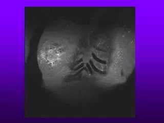
  14. Breast Specimen Radiography Excisional Biopsy Mammotome or FNAB specimen

  15. Pathologies

  16. Types of Pathologies • Cyst • Lipoma • Fibroadenoma • Fibrocystic Breasts • Cancer • DCIS – Ductal Carcinoma in Situ • IDC – Infiltrating Ductal Cancer

  17. Cysts

  18. Cysts

  19. Lipoma

  20. Fibroadenoma

  21. Fibroadenoma

  22. Fibrocystic breasts

  23. Cancer

  24. Cancer

  25. Breast Calcifications

  26. Breast Calcifications

  27. DCIS – Ductal Carcinoma in Situ

  28. DCIS – Ductal Carcinoma in Situ

  29. Infiltrating Ductal Cancer

  30. Infiltrating Ductal Cancer

  31. Other Invasive Breast Procedures

  32. Galactography / Ductography

  33. Galactography / Ductography

  34. Indications of Galactography • Nipple Discharge • White / Yellow/ Green / Brown / Red • Can be considered benign or malignant • Approx 2-5% bloody discharges = cancer • Other causes can be a blocked duct due to a papilloma (shows as a filling defect on film) • Spontaneous discharge more worrisome than if discharge must be expressed manually

  35. Galactography / Ductography • Filling defect • Could be an indication of ductal papillomas

  36. Other Imaging Modalities

  37. Ultrasound Breast

  38. Breast MRI

  39. MRI vs Mammo

  40. MRI vs. Mammo

  41. Digital vs. Conventional

  42. Digital vs. Conventional

More Related