Download
1 / 20
200 likes | 335 Views
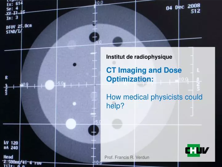
CT Imaging and Dose Optimization: How medical physicists could help?. Prof. Francis R. Verdun. Summary. Medical physicists and CT dose optimization ? Context of our experience Results obtained from last year In search to be useful in patient dose optimization framework

E N D
CT Imaging and Dose Optimization:How medical physicists could help? Prof. Francis R. Verdun
Summary • Medical physicists and CT dose optimization ? • Context of our experience • Results obtained from last year • In search to be useful in patient dose optimization framework • What has been learned • Where are we going to put our efforts
Quality assurance in Switzerland before 2010 • Manufacturers are authorized by FOPH to: • Installand test the system before the 1st patient • Check a list of IECrequirements • Do the preventive maintenances • FOPH organizes audits in centers • Advantages • Simple, relatively homogeneous, in a way, cost efficient • Main limitations • Performances are detector oriented (not patient oriented) • Manufacturers are both judge and judged • Does not comply with Euratom 97/43 requirements
Towards legal compliance Certainly not perfect but a decent working basis 2011 : Working group proposes a concept “Requirements for medical physicists in Nuclear Medicine and Radiology” (June 2011)”
Outcomes from our 2013 experience • 45 units tested • About15% of the CT units in use in Switzerland • Problems found • QC: • 4 CT : Laser poorly adjusted • 21 CT : Hounsfield unit not calibrated if kV ≠ 120 • Use of the unit: • Tube current modulation is not used optimally • Penumbra and over-ranging phenomena are poorly handled • Dose Reference Level often misunderstood
Use of the unit on patient Mean dosimetric results for CT examinations. The CTDI and DLP are given per phase, whereas E is given per examination Minimum and maximum patient effective doses according to ICRP 103. All values are given per examination
Proposal for 2014 • Sampling of examinations • Use common indications • A more practical QC evaluation • Use of an objective way to assess low contrast resolution • Assess the actual benefit of iterative recons • Benchmark a few clinical protocols • Be more on the image quality side than dose
Focus on the way the CT unit is used • Choice of protocol indications • Head • Trauma • Sinus • Willis polygon • Chest • Pulmonary embolism suspected • Seek for a primary tumor • Abdomen • UroCT • Seek for a primary liver tumor
Challenges to CT image evaluation • CT systems are not linear and not shift-invariant • Resolution/sharpness depends on object size, contrast, location, and noise level • There isn’t one MTF that describes the system transfer • Use of the concept of the Task Transfer Function (TTF) • Noise is colored • Pixel variance doesn’t tell the whole story • Noise is non-stationary • Noise texture or NPS depends on location • Iterative reconstruction introduces more difficulties
Task Transfer Function Phys Med Biol. 2014 Aug 7;59(15):4047-64
Main challenge • Dose reduction and loss of low contrast resolution • Iterative reconstructions remove some signs of high noise level • One can have a reasonable looking image while low contrast structures are not transferred • Use of an objective way to assess low contrast resolution
Observer’s template Image Principle of linear model observers The LMO computes the decision variable l for each image
Image noise Image with signal x x = = Response ls Response ln
ROC curve construction From the values of ln and ls : observer response Comparison with human observers Area under Curve : AUC dA
Expected outcomes • Ensure that dose reduction reduces low contrast resolution in a safe way • Adequacy of the protocol with the level of image quality required • Iterative is not a magic solution, radiologists need to know what is lost and judge if it is OK or not • Promote the use of new technological progresses while being critic
Conclusion • How medical could help in CT optimization? • Focus on image quality assessments in CT • We should never speak about dose without noticing image quality • Help radiologists in setting minimum low contrast resolution requirements • Protocol optimization indication based • Ideally it would be nice to link a dose report to an image quality performance
Thankyou for your attention Acknowledgements Nick Ryckx, Julien Ott, Damien Racine