Download
1 / 47
690 likes | 1.46k Views
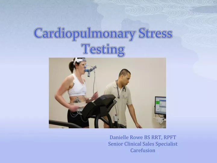
Cardiopulmonary Stress Testing. Danielle Rowe BS RRT, RPFT Senior Clinical Sales Specialist Carefusion. WHAT IS THE PURPOSE OF CPX TESTING?. It is an assessment of a subject’s functional (work) capacity under stress. We judge this functional capacity by primarily evaluating peak VO2.

E N D
Cardiopulmonary Stress Testing Danielle Rowe BS RRT, RPFT Senior Clinical Sales Specialist Carefusion
WHAT IS THE PURPOSE OF CPX TESTING? • It is an assessment of a subject’s functional (work) capacity under stress. • We judge this functional capacity by primarily evaluating peak VO2. • After functional capacity is established, we then are provided with insight on cause of limitations if they exist.
CO2 O2 Possible limitations to Exercise • Ventilation • Gas Exchange • Circulation (inc. ECG) • Metabolism metabolism Peripheral Gas Exchange Pulmonary Gas Exchange
Indications For Cardiopulmonary Exercise Testing • Evaluation of exercise tolerance • Evaluation of undiagnosed exercise intolerance • Evaluation of patients with cardiac disease • Evaluation of patients with Respiratory disease • Specific Clinical Applications • Preop assessment on certain populations, rehab evaluations, disability, transplantation etc. From ATS/ACCP
Max or Submax Testing • For Cardiopulmonary diagnostic purposes, MAXIMAL testing is performed. • We aim to stress the cardio-respiratory system until we identify the factor which limits exercise capacity. • Sub max test are more common in athletics training, rehabilitation etc.
Exercise Protocols • Cycle Ergometry • Ramp, typically increases of 5 -30 W/minute • Aim to have exercise portion of testing lasting approx. 10 minutes • 1-3 minutes resting data • 1-3 minutes unloaded pedaling • Treadmill • Speed constant and grade increased (Balke Protocol). 2 mph, 0% grade and then the grade is increased 2-3% every minute. • Speed and grade are both increased ( Bruce Protocol). 1.7 mph, 10% grade and then increased by .8 mph and 2% grade every 3 minutes.
Bike vs Treadmill Bike Treadmill • VO2 Max lower higher • Work Rate Meas. Yes No • Blood gas collection easier harder • Noise and artifacts less more • Safety safer less safe? • Wt. bearing in obese less more • Leg muscle fatigue often less
Bike vs. Treadmill cont. • Bike • All things considered, bike is considered the most appropriate for patients. • This is due to ability to accurately quantify work and the minimizing of test artifacts. • Just remember vigorous encouragement is often needed near peak of exercise test to help overcome and push through leg muscle fatigue. • Treadmill • VO2 max is often 5-10% higher • Most appropriate for athletes and patients in whom abnormalities may occur only with the highest demand ( cardiac ischemia).
Selecting the Work Rate 5 Watts/min Severe impairment (e.g. patient who is confined to home or walks only short distances) 10 Watts/min Moderate impairment (e.g. patient who walks one or two city blocks before symptoms) 15 Watts/min Mild impairment or sedentary older patient 20 Watts/min Sedentary younger patient 25 Watts/min Active younger patient (regular sports, physical exercise) 30 Watts/min Athletic and fit (competitive sports) 40 Watts/min Extremely fit (highly competitive) Chris Cooper, MD.. Harbor UCLA Medical
Was it a Good Test ? • How can we tell ? • Most common problem seen is sub-maximal performance. • Patients are supposed to “suffer” - that is the whole idea of the test - to put them under stress. • Don’t stop when the patient hits Max Predicted Heart Rate - carry on until the patient stops.
Often the best Indicator.. • RER • “Although no one RER value defines maximal effort, values greater than 1.15 are more likely to be associated with near maximal or maximal effort.” From ATS/ACCP Statement
RER (or RQ) = VCO2/VO2 • At Baseline, RER < 0.8 • If it isn’t, check for hyperventilation • If no hyperventilation, something is wrong • Many patients hyperventilate in the baseline state • At end of test, RER > 1.15 • Indicates maximal exercise effort and therefore a good test
Indications for Exercise Termination • Chest pain suggestive of ischemia • Ischemic ECG changes • Complex ectopy • Second or third degree heart block • Fall in systolic pressure >20 mmHg from the highest value during the test. • Hypertension ( >250 mmHg sys; >120 mmHg diastolic) • Severe desaturation: SpO2 ≤ 80% when accompanied by symptoms and signs of severe hypoxemia • Sudden pallor • Loss of confusion • Dizziness or faintness • Signs of respiratory failure From ATS/ACCP Statement
VO2 Peak/Max • Maximal Oxygen Uptake (VO2 max) • Represents the highest VO2 that can be reached as evidenced by a failure for VO2 to increase further despite and increase in work rate. • Peak Oxygen Uptake ( VO2 peak) • Represents the highest VO2 reached during the test where a presumed maximal effort was given. • These Terms are often used interchangeably.
Oxygen Consumption • The amount of oxygen used per minute. • 250 mL/min at rest (3.5 mL/min/kg) • 5,000 mL/min at strenuous exercise (>70 mL/min/kg) Expected Vo2 mL/min/kg values from American Heart Association
Peak VO2 Cont. • Peak VO2 is often used in the course of treating heart failure and in selecting heart transplant candidates: • Typically these subjects that test with a peak VO2 <14 ml/min/kg are strongly considered for transplantation. • Peak Vo2 is often considered with major surgery especially abdominal surgery in elderly or sick patients. • >20 ml/min/kg good prognosis • <15 ml/min/kg high risk
VO2- Work relationship • VO2/Work slope = Normal is right around 10/ml/min/watt with relatively small range of normal reported in many studies. Typically (8.5- 11) considered normal. • Shallower slope, so a value < 8.5- 8.7 ml/min/watt point to a problem of O2 flow or O2 utilization.
Anaerobic Threshold (AT) • Lactacte Threshold, Lactic Acid Threshold, Gas Exchange threshold, Ventilatory threshold etc.. • The highest level of oxygen consumption that can be sustained without developing metabolic acidosis. • The point at which anaerobic metabolism starts to contribute.
AT cont. • Normally occurs at about 50-60% of Vo2 max predicted however there is a wide range of normal reported (35-80%). • 40% is the generally accepted lower limit of normal used clinically.
Detecting ATInvasive determination • Arterial Lactate measurements. AT is graphically determined by plotting lactate concentration vs. Vo2.
AT summary • Athletes can do more activity aerobically so AT is increased. • In diseases ( most cardiovascular) that affect O2 supply to exercising muscles , the AT is often found to be early. This can also occur in more rare conditions with mitochondrial myopathies. • In clinical CPX testing you are searching for an early AT. If it is not early, then it really does not matter precisely where it occurred.
Cardiac Parameters • Heart Rate and HR reserve • BP <220/90 • O2 pulse • ECG • HR –VO2 relationship
Heart Rate and HR reserve • Many formulas exist for predicting max HR. • 220- age • 210- (age × 0.65) • HR reserve is the difference between the predicted max and the achieved max. • Normally there is very little to no reserve in normal subjects giving maximal effort. • Often a HRR < 15 beats/min is normal.
HR- VO2 Relationship 200 - Predicted Maximum Heart Rate HRR = 0 HR (b/min) 0 | 0 VO2 Max Predicted HR/VO2 normal = 3-4 beats/ml/min/kg
O2 pulse • VO2/HR • Amount of O2 uptake per each beat of heart. • Dependent on stroke volume and O2 uptake. • O2 pulse = SV × C(a-v)O2 • O2 pulse normally increases with incremental exercise due to increases in both SV and O2 extraction. • When O2 content and C(a-v)O2 are maximal and assumed to be normal ( approx. 15 ml/dl), stroke volume can be estimated: • SV= O2 pulse/15 *100
Ventilatory Parameters • VE • RR • Tidal volume • Breathing reserve • Inspiratory capacity trending ( Ex. FVL) • Capacity, reserve assessment
VE and Breathing reserve • Measure Spirometry and MVV prior to exercise test. Then decide which to use as predictor of maximal ventilation. Quality of these baseline measurements is key….be careful! • Often MVV may not be the best indicator of capacity as breathing pattern of the 12-15 sec effort is not typically a pattern subject adopts during exercise. • FEV1 × 35 or 40 is often used to estimate MVV or capacity. • Things to consider: • Effort and quality of baseline spirometry and MVV. Can I use either? • It may be better to use actual MVV in cases of upper airway obstruction or neuromuscular weakness. • Breathing reserve= (MVV-VE max/ MVV)× 100 • Normal = 20-30 %. Typically < 15% considered low.
Breathing pattern • The rise in VE during exercise is associated with an increase in both depth and frequency of breathing. • Tidal Volume- Vt typically increases until it reaches about 50-60% of VC or 70% of IC. • Further increases in VE are accomplished by respiratory rate. • Typically the rate does not normally exceed around 55 bpm.
Ventilatory Equivalents • How many liters of air we need to breath to exchange 1 L of gas. • VEVO2 • VECO2 • Indicators of efficiency of ventilation. • Increases in ventilatory equivalents: • Often found in diseases in which pulmonary blood flow is abnormally reduced to ventilated gas exchange units.
Ventilatory Equivalents (Efficiency of ventilation) 40 VE/CO2 VE/VO2 20 RC AT 0 VE/VO2 @ AT: 26.5 (22.1 – 30.9) VE/VCO2 @ AT: 29.1 (24.8 – 33.4) NORMAL VALUES: Ref: Wasserman
Arterial Blood SamplingDo we need it? • Sometimes…. • When the adequacy of pulmonary gas exchange is in question up front. Typically in diseases like ILD, pulmonary vascular disease, COPD with low DLCO etc. • When concern over increased dead space is and issue. You want real VD/VT measurements. • When patients have an abnormal initial CPET maybe showing increased VE/VCO2 but no specific reason. Was it hyperventilation or due to increased VD. • If you do it, do it right! Use and Arterial line. No single samples at peak exercise.
Additional pieces of information when blood gas sampling is done • Information about ability to exchange oxygen. • PaO2- normally >80 mmHg and should not decrease with exercise. • P (A-a)O2- should be <35 mmHg at peak. • SaO2
ABG cont.. • VD/VT = (PaCO2 – PECO2)/PaCO2 • Fraction of each breath “wasted” on ventilating anatomic and physiologic dead space. • You need PaCO2 to get a true VD/VT. You cannot accurately estimate! • The estimated VD/VT that is often reported uses end-tidal PCO2 in place of PaCO2 which can be misleading. • Normally PetCo2 is a little less than PaCO2 at rest but becomes greater than PaCO2 with exercise leading to an overestimation of VD/VT. • With lung disease PetCO2 may remain below PaCO2 even with exercise causing an underestimation of dead space.
VD/VT • Normal at rest is 30-40%. • Should fall with exercise due to increasing tidal volume. • Typically at peak exercise we should see values less than 28% in subjects < 40 yrs. Values less than 30% normal in subjects >40 yrs.
Putting it all together • Was it a good test? • Can subject achieve normal VO2 and do normal amount of “work”? • Is there a premature metabolic acidosis? • Low AT • Is there a cardiovascular limitation? • Is there a ventilatory (mechanics) limitation? • Does pulmonary gas exchange contribute to exercise limitation?
Obesity • High O2 cost to perform work. • Peak VO2/kg is low when expressed per kg of actual wt but normal when expressed per kg of ideal wt. • Low PaO2 that often normalizes with exercise. • Failure to develop normal ventilatory compensation for metabolic acidosis.
Cardiovascular disease • Low VO2 • Early AT • Reduced maximal O2 pulse • Steep HR/VO2 relationship • Peak HR variable- may be normal or reduced. • Often increased ventilatory reserve
Ventilatory limitation • Low VO2 • High VD/VT • Low breathing reserve • High heart rate reserve • AT normal or not reached • Failure to develop respiratory compensation for metabolic acidosis.