Download
1 / 70
740 likes | 1.49k Views
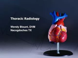
Thoracic Anatomy & Physiology A Simple Review. Mark Welliver CRNA, MS Assistant Professor. Diagram of Thoracic Area. The Larynx. epiglottis. hyoid bone. thyroid cartilage. cricoid cartilage. trachea. Trachea & Bronchi. Lung Anterior. Lung Posterior. Lung Left Side. Lung Right Side.

E N D
Thoracic Anatomy & PhysiologyA Simple Review Mark Welliver CRNA, MS Assistant Professor
The Larynx epiglottis hyoid bone thyroid cartilage cricoid cartilage trachea
Dynamics of pulmonary blood flow • Blood flow is greatest in dependent parts of lung- gravity • Hypoxic Pulmonary Vasoconstriction (HPV) redistributes blood away from poorly ventilated alveoli
Spontaneous ventilation Perfusion greatest at bases
Remember- Blood flow greatest at bases- Dependant Area Gravity pulls blood flow to bases
Dynamics of Spontaneous Breathing • Diaphragm descends causing a negative intrathoracic pressure • Gas flows from higher pressure to lower pressure • Greatest gas flow in spontaneous ventilation is to bases
Spontaneous ventilation Ventilation greatest at bases
Dynamics of Spontaneous Breathing • Apex alveoli already distended from greater NEGATIVE pleural pressure thus they have less compliance to expand and receive volume increases • Apex ribs short and expand minimally • Base alveoli have greatest gas flow due to greater change in thoracic pressures during insp.- exp. phases d/t insp. diaphragmatic downward movement d/t pale handle effect • Abdominal contents pushing up and gravity pulling lungs down lessens the negative pleural pressure in bases
CHEST WALL PLEURAL SPACE * *Greater negative pressure in apex during end expiration- small change during inspiration pale handle effect lung follows LUNG diaphragm moves down
Pale handle effect • Internal intercostals, pull downward, aid expiration • External intercostal, elevate ribs, aid inspiration. • Pneumonic; In-Ex, Ex-In
Intercostals Note; internal and external intercostal muscles
Lungs want to recoil, Thoracic cage wants to expand • Thus, the pleural cavity has a vacuum ( a negative pressure)
Spontaneous ventilation • Ventilation(V) to Perfusion(Q) well matched in spontaneous ventilating patients • Decreasing intra-pleural pressure during inspiration draws inspired gas into bases of lung where there is the most blood flow • Pleural pressure end exp. –5 cm H2O • Pleural pressure during insp. –7.5 H2O • Pleural pressure change 2.5 cm H2O
Thoracic Pressure Differences • Driving pressure- Pressure difference between two points in a tube or vessel(force) • Trans airway pressure-Barometric pressure difference between the mouth pressure and alveolar pressure • Trans pulmonary pressure- The pressure difference between alveolar pressure and pleural pressure • Trans thoracic pressure- The difference between alveolar pressure and the body surface pressure • Pleural pressure- The primarily negative pressure in the pleura
Changes in lung volume, alveolar pressure, pleural pressure, and trans pulmonary pressure during normal breathing
Ventilation/Perfusion V/Q • Ventilation is closely matched to perfusion • Normal V/Q matching is 0.8 • Causes of mismatching include; physiologic shunt hypoventilation disease states.......
Pressure Dynamics within lung units: Alveolar (A) arterial (a) venous (v)
Zones of West PA>Pa>Pv 1 Pa>PA>Pv 2 Pa>Pv>PA 3
Zone 1 Alveolar pressure exceeds arterial exceeds venous A a v
Zone 2 Arterial pressure exceeds Alveolar exceeds venous A v a
Zone 3 Arterial pressure exceeds venous exceeds Alveolar A v a
Zones of West Alveoli Volume representation of end expiration to end inspiration
Mechanical ventilation Greatest blood flow to bases Greatest gas flow to apexes
Mechanical ventilation Greatest gas flow to apexes of lung
Mechanical ventilation • Ventilation(V) to Perfusion(Q) poorly matched in mechanically ventilated patients • Positive pressure ventilation pushes gas into apexes of lung. Path of least resistance. Blood perfuses primarily the dependant parts of lung again due in part to the pull of gravity
Hypoxic Pulmonary Vasoconstriction HPV • HPV effectively redirects blood flow away from hypoxic or poorly ventilated lung units • Pulmonary vascular endothelium release potent vasoconstrictor peptides called endothelins • Volatile anesthetics above 1 mac and nitrous oxide block HPV
Mechanical ventilation • Gas flow to apex and blood flow to bases= V/Q mismatching • Poorly ventilated alveoli are prone to atelectasis and collapse
Atelectasis • Atelectasis is essentially collapse of pulmonary tissue that prevents O2 & CO2 exchange. • Primary causes: obstruction of airway and lack of surfactant • Absorption atelectasis is caused by occlusion of an airway with resultant absorption of trapped gas and collapse of alveoli. higher [O2] worsens due to removal of N as an inert stabilizer • Hypoventilation during positive pressure ventilation is often primary cause of absorption atelectasis
Review • General anesthetics above 1 mac block HPV • Mechanical ventilation alters gas flow dynamics • Paralysis increases resistance to gas flow • Absorption atelectasis frequently seen to varying degrees
Worsening V/Q mismatch numeric representation ONLY not actual values. Causes?
Open Chest Ventilation Dynamics • Paradoxical ventilation • Closed (simple) pneumothorax • Communicating pneumothorax • Tension pneumothorax • Hemothorax
Closed(simple) pneumothorax • No atmospheric communication • Treatment based on size and severity-catheter aspiration, thoracostomy, observation
Communicating pneumothorax “sucking chest wound” • Affected lung collapses on inspiration and slightly expands on expiration • Treatment: O2,thoracostomy tube, intubation, mech. vent.
Tension pneumothorax • Air progressively accumulates under pressure within pleural cavity. Compressing other lung, great vessels • Treatment; immediate needle decompression
Hemothorax • Accumulation of blood in pleural space • Treatment; airway management,support hemodynamics, evacuation
Lung Isolation Tubes/ Techniques • Single-Lumen Endobronchial Tubes • Endobronchial Blockers • Double-Lumen Endobronchial Tubes See also Lung Isolation Tutorial Power Point