Download
1 / 86
870 likes | 890 Views
Chapter 9 Motor System - 2. Spinal Reflex, Descending Pathways and Cerebellum. Content. Spinal Reflexes Function of Brain Stem Function of Cerebellum. Reference. P482-489 P493-496. UNIT XI The Nervous System: C. Motor and Integrative Neurophysiology. P164 - 180.

E N D
Chapter 9 Motor System - 2 Spinal Reflex, Descending Pathways and Cerebellum
Content • Spinal Reflexes • Function of Brain Stem • Function of Cerebellum
Reference P482-489 P493-496 UNIT XI The Nervous System: C. Motor and Integrative Neurophysiology P164 - 180
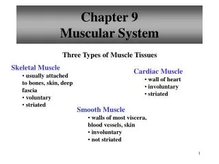
Section 1. Spinal Reflexes • Somatic reflexes mediated by the spinal cord • May occur without the involvement of higher brain centers • Was facilitated or inhibited by brain • For example • Stretch reflex • Deep tendon reflex • Crossed extensor reflex • Superficial reflex
1 Anatomy of Muscle Spindle • 3-10 intrafusal muscle fibers • detect change in the length of the muscle -- stretch receptors that report the stretching of the muscle to the spine. • The central region and peripheral region of the intrafusal fibers
Anatomy of Muscle Spindle • Intrafusal fibers are wrapped by two types of afferent endings • Primary sensory endings • Type Ia fibers • Innervate the center of the spindle • Secondary sensory endings • Type II fibers • Associated with the ends of the nuclear chain fiber
Components of muscle spindle Dynamic intrafusal fiber Static intrafusal fibers Static intrafusal fibers Primary ending } Ia Afferent axons II Secondary ending } Nuclear Bag Fiber Nuclear Chain Fiber
Anatomy of Muscle Spindle • Primary sensory endings • Type Ia fibers • Stimulated by both the rate and amount of stretch (dynamic response)
Anatomy of Muscle Spindle • Secondary sensory endings • Type II fibers • stimulated only by degree of stretch (static response)
Anatomy of Muscle Spindle • The contractile region of the intrafusal muscle fibers are limited to their ends • only these areas contain actin and myosin filaments • are innervated by gamma () efferent fibers
Muscle stretch reflex Definition: Whenever a muscle is stretched, excitation of the spindles causes reflexive contraction of the same muscle from which the signal originated and also of closely allied synergistic muscle. The basic circuit: Spindle Ia or II nerve fiber dorsal root of the spinal cord synapses with anterior motor neurons -motor N. F. the same M. from whence the M. spindle fiber originated.
The Stretch Reflex • Exciting a muscle spindle occurs in two ways • Applying a force that lengthens the entire muscle • Activating the motor neurons that stimulate the distal ends of the intrafusal fibers to contact, • thus stretching the mid-portion of the spindle (internal stretch)
The Stretch Reflex • Whatever the stimulus, when the spindles are activated • their associated sensory neurons transmit impulses at a higher frequency to the spinal cord
The Stretch Reflex • The reflexive muscle contraction that follows resists further stretching of the muscle
The Stretch Reflex • Branches of the afferent fibers also synapse with inter- neurons that inhibit motor neurons controlling the antagonistic muscles
Inhibition of the antagonistic muscles is called reciprocal inhibition • causes the antagonists to relax
The types of the Stretch Flex Tendon reflex (dynamic stretch reflex) Caused by rapid stretch of the muscle, as knee-jerk reflex; Transmitted from the IA sensory ending of the M. S. Causes an instantaneous, strong reflexive contraction of the same muscle; Opposing sudden changes in length of the M.; • A monosynaptic pathway • being over within 0.7 ms;
The types of the Stretch Flex 2)Muscle tonus (static stretch reflex): Caused by a weaker and continues stretch of the muscle, Transmitted from the IA and II sensory ending of the M. S. Multiple synaptic pathway, continues for a prolonged period. Non-synchronized contraction, M. C. for at least many seconds or minutes, maintaining the posture of the body.
The Stretch Reflex • most important in large extensor muscles which sustain upright posture • Contractions of the postural muscles of the spine are almost continuously regulated by stretch reflexes (Muscle Tonus )
When a muscle is suddenly stretched, a signal is transmitted over Ia sensory fibers from muscle spindles. It will induces Contraction of the muscle in which the active spindles are located Relaxation of the muscle in which the active spindles are located Contraction of muscles antagonistic to those in which the active spindles are located Relaxation of intrafusal fibers in the active spindles Direct synaptic activation of gamma motor neurons
Muscle spindle: motor innervation • Gamma motoneurons: • Innervate the poles of the fibers.
Descending influence (UMN) 1a a g-LOOP Muscle spindle g Activation of the g-loop results in increased muscle tone MUSCLE
Functional significance of gamma impact on spindle activity • The tension of intrafusal fibers is maintained during active contraction by gamma activity. • The system is informed about very small changes in muscle length.
Which of the following statements about muscle and passive stretch of muscle spindles is true? Primary (Ia) sensory fibers increase their firing rate Secondary sensory fibers decrease their firing rate Alpha motor neurons are inhibited Gamma motor neurons are stimulated Muscle spindles go completely slack
Structure and Innervation of Golgi Organ • Located in the muscle tendon junction. • Connective tissue encapsulating collagen fibers and nerve endings. • Attached to 10-20 muscle fibers and several MUs. • Ib afferent fiber. • sensitive to tension
Golgi tendon organ: response properties • Less frequent than muscle spindle • Sensitive to the change of tension caused by the passive stretch or active contraction
The Deep Tendon Reflex • When muscle tension increases moderately during muscle contraction or passive stretching, • GTO receptors are activated and afferent impulses are transmitted to the spinal cord
The Deep Tendon Reflex • motor neurons in the spinal cord supplying the contracting muscle are inhibited and antagonistic muscle are activated
The Deep Tendon Reflex • cause muscle relaxation and lengthening in response to the muscle’s contraction • opposite of those elicited by stretch reflexes • help ensure smooth onset and termination of muscle contraction • important in activities involving rapid switching between flexion and extension such as in running
In the patellar tendon reflex, which of the following items will synapse directly on alpha motor neurons that innervate the muscle being stretched? Ia sensory fiber Ib sensory fiber Excitatory interneurons Gamma motor neurons Inhibitory interneurons
Part 3. The Crossed Extensor Reflex • The reflex occur when you step on a sharp object • There is a rapid lifting of the affected foot (ipsilateral withdrawal reflex ), • while the contralateral response activates the extensor muscles of the opposite leg (contralateral extensor reflex) • support the weight shifted to it
Part 4. Superficial Reflexes • elicited by gentle cutaneous stimulation • dependent upon functional upper motor pathways • Babinski reflex
Babinski reflex - an UMN sign • Adult response - plantar flexion of the big toe and adduction of the smaller toes • Pathological (Infant) response - dorsoflexion (extension) of the big toe and fanning of the other toes • Indicative of upper motor neuron damage
The withdrawal reflex is initiated by stimulation delivered to which of the following receptors? Muscle spindle Joint capsule receptor Cutaneous free nerve ending Golgi tendon organ Pacinian corpuscle
Part 5. Spinal cord transection and spinal shock • Concept:When the spinal cord is suddenly transected in the upper neck, essentially all cord functions, including the cord reflexes, immediately become depressed to the point of total silence. (spinal animal)
(2) During spinal shock: complete loss of all reflexes, no tone, paralysis complete anesthesia, no peristalsis, bladder and rectal reflexes absent (no defecation and micturition ) no sweating arterial blood Pressure decrease(40mmHg)
(3) the reason:The normal activity of the spinal cord neurons depends on continual tonic excitation from higher centers (the reticulospinal-, vestibulospinal- corticospinal tracts). (4) The recovery of spinal neurons excitability.
Section II. Role of the brain stem Support of the Body Against Gravity – Roles of the Reticular and Vestibular nuclei