Download
1 / 1
10 likes | 419 Views
A. B. A. B. C. D. C. D. F. E. No. 099. PENILE TRACTION AND PEYRONIE’S DISEASE: IN-VITRO ANALYSIS OF THE EFFICACY OF MECHANICAL TRACTION ON CELLULAR CHANGES IN PEYRONIE ’ S PLAQUE IN A STRAIN CULTURE SYSTEM.

E N D
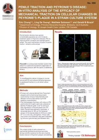
A B A B C D C D F E No. 099 PENILE TRACTION AND PEYRONIE’S DISEASE: IN-VITRO ANALYSIS OF THE EFFICACY OF MECHANICAL TRACTION ON CELLULAR CHANGES IN PEYRONIE’S PLAQUE IN A STRAIN CULTURE SYSTEM Eric Chung1,2,, Ling De Young2, Matthew Solomon1,2 and Gerald B Brock2 1 Department of Urology, Princess Alexandra Hospital, Brisbane, QLD Australia 2 Division of Urology, St Joseph Health Care, London, ON Canada Introduction Penile traction devices have gained considerable popularity as a non-invasive treatment option in Peyronie’s disease (PD) to improve penile curvature and maintain penile length. The exact mechanism of action of penile traction remains controversial. Flexcell® is a pressurised chamber system that allows for sustained and dynamic application of hydrostatic pressure with the option of additional tension. Results Figure 1: Representative images from control (A,C,E) and PD (B,D,F) Primary cell culture magnification x400. A&B Hsp47, C&D TGF β1Receptor. E&F Fibronectin (green) and α-actin (red). Increased TGF β1 receptor and α-actin expression was evident in PD cells. Aim To investigate the cellular changes to normal tunical and PD cell cultures following exposure to equibiaxial traction in a cell-strain culture system. Figure 2: Data presented mean ± SE of 6 control (colored bars) and 10 PD cell protein by Western blot densitometric analysis. Significant increase in smooth muscle α-actin, β-catenin and Hsp 47 proteins was detected in the PD group. • Methods • Primary cell cultures derived from Peyronie’s plaques and normal tunical tissue were examined by immuno-histochemistry and Western immuno-blot assay to provide baseline characteristics. • The first passage of primary cell culture of PD and Control (3 of each) were sub-cultured on BioFlex-ProNectin plate 1x10⁶ for 24 hours then exposed to a Flexcell Strain Unit (FX 5000, Flexcell International) to generate a sinusoidal strain of 18% at 1Hz (strained group) on the biaxial plates for 24 hours under 37° C in a 5% CO2 atmosphere. Those cells and control (non-strained) cells were evaluated by immuno-cytochemistry and Western blot. GADPH PD Control SControl SPD MMP-8 β-catenin MMP-8 Figure 3: Data presented mean ± SE of 3 PD and control compare with strained (colored bars) cell protein by Western blot densitometric analysis. IncreasedMMP-8 expression in SPD group was detected. There was no measured change in β-catenin level. Conclusions This unique study of cells cultured in a mechanical strained environment provides strong scientific evidence for the use of penile traction device in Peyronie’s plaque remodelling. Figure 4: Representative images from PD (A) and SPD (B) of a actin (arrow) magnification x200, magnification x400 (C) and (D). Decreased α-actin staining post-stretch was detected. Poster presentation sponsor