Download
1 / 24

E N D
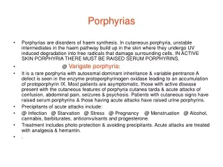
Porphyrias • Porphyrias are disorders of haem synthesis. In cutaneous porphyria, unstable intermediates in the haem pathway build up in the skin where they undergo UV induced degradation into free radicals that damage surrounding cells. IN ACTIVE SKIN PORPHYRIA THERE MUST BE RAISED SERUM PORPHYRINS. • @ Varigate porphyria: • It is a rare porphyria with autosomal dominant inheritance & variable pentrance A defect is seen in the enzyme protoporphyrinogen oxidase leading to an accumulation of protoporphyrin IX. Most patients are asymptomatic, those with active disease present with the cutaneous features of porphyria cutanea tarda & acute attacks of confusion, abdominal pain, seizures & psychosis. Patients with cutaneous signs have raised serum porphyrins & those having acute attacks have raised urine porphyrins. • Precipitants of acute attacks include: • @ Infection @ Starvation @ Stress @ Pregnancy @ Menstruation @ Alcohol, cannabis, barbiturates, anticonvulsants and progesterone. • Treatment includes photo protection & avoiding precipitants. Acute attacks are treated with analgesia & hemantin. • .
Varigate porphyria
Porphyrias (continue) • Erythropoietic protoporphyria(EPP) • It is due to the impaired activity of the enzyme ferrochelatase. This result in accumulation of its substrate protoporphyrin. Protoporphyrins in the skin are excited by light causing local photoxidative damage & symptoms such as immediate tingling, itching, erythema & purpura.Protoporphyrins also ACCUMULATE IN LIVER HEPATIC DAMAGE & ARE EXCRETED IN THE BILE PREDISPOSING TO GALLSTONES. EPP usually presents in children & has an equal sex distribution, both autosomal dominant & autosomal recessive inheritance have been reported. With time patients develop pitted scars & thickened skin on the cheeks, nose & dorsum of the hand. • Treatment is with photoprotection, beta carotene, cysteine & cholestyramine. In the majority of patients, the cutaneous features are mild, only a small umber of patients develop significant liver disease.
The amount of porphobilinogen (PBG) in urine is increased during attacks of AIP. There are three acute porphyrias that can cause increases in PBG, namely AIP, Hereditary Coproporphyria (HCP) and Variegate Porphyria (VP). Acute attacks can occur in all of these conditions. Skin photosensitivity can occur in HCP and VP, but not AIP. Urine may appear purple during an attack or after standing in light
Typical cutaneous lesions in a patient with porphyria cutanea tarda.
Porphyria cutanea tarda Subepidermal bulla formation has resulted in loss of the epidermis. Note the rigid papillary dermal capillary walls. Porphyria cutanea tarda
Erythropoietic protoporphyria
Cutaneous histologicalfeatures of EPP with PAS positive material deposition around papilar blood vessels and dermo epidermal junction..
Erythropoietic Protoporphyria • Erythema and edema of the hands
Yellowish coloration of the teeth of a child with congenital erythropoietic porphyria. B, Red fluorescence (erythrodontia) can be readily observed under Wood light. Actas DermaoSifilo 1O4: 212, 2O13
Erythropoietic protoporphyria pathology In the dermis eosinophilic hyaline material is deposited in and around blood vessel walls. The deposition can be extensive and involve the surrounding dermis to mimic a colloid milium . Impressive thickening of vessels is seen at higher power examination
Erythropoietic protoporphyria Gallstones