Download
1 / 65
760 likes | 2.17k Views
JAUNDICE IN PEDIATRICS. DENNIS S. FLORES, MD, DPPS, DPSN, DPNSP. SCOPE. Definition of jaundice Bilirubin metabolism Mechanisms of jaundice Physiologic vs pathologic jaundice Breastfeeding vs breastmilk jaundice Management of neonatal jaundice

E N D
JAUNDICE IN PEDIATRICS DENNIS S. FLORES, MD, DPPS, DPSN, DPNSP
SCOPE • Definition of jaundice • Bilirubin metabolism • Mechanisms of jaundice • Physiologic vs pathologic jaundice • Breastfeeding vs breastmilk jaundice • Management of neonatal jaundice • Cholestasis (neonatal hepatitis vs biliary atresia) • Management of cholestasis • Hepatitis A and B virus infections/vaccination
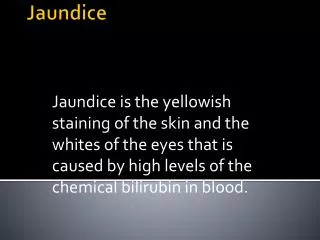
JAUNDICE • Yellowish discoloration of the sclera, skin, and mucous membranes • Clinically apparent when bilirubin level reaches 2-3 mg/dl in children and adults, and >5 mg/dl in neonates
JAUNDICE • May be physiologic • It may be the earliest and only sign of hepatic dysfunction • Liver disease must be suspected in the infant who appears only mildly jaundiced but has dark urine or acholic stools (light colored)
BILIRUBIN • Comes in 4 forms: • Unconjugated bilirubin • Tightly bound to albumin • Free or unbound bilirubin • The form responsible for kernicterus, because it can cross cell membranes • Conjugated bilirubin • The only fraction to appear in urine • Delta fraction • Bilirubin covalently bound to albumin
BILIRUBIN • DIRECT BILIRUBIN • Conjugated bilirubin and delta bilirubin • INDIRECT BILIRUBIN • Unconjugated bilirubin
BILIRUBIN • Is a bile pigment produced by the breakdown of heme and reduction of biliverdin • Unconjugated bilirubin is insoluble in plasma unless bound to protein, mainly albumin • Normally, 95% of the circulating bilirubin is unconjugated
BILIRUBIN • Bilirubin-albumin complex is dissociated by receptors on hepatocytes • Albumin – remains in the plasma • Bilirubin – taken into the hepatocyte and conjugated by the enzyme bilirubin UDP-glucuronyl transferase to form bilirubin diglucuronide, which is excreted into the biliary system
BILIRUBIN • In the gut (colon), bilirubin diglucuronides are degraded by bacteria and converted into a mixture of compounds, known as urobilinogen and stercobilinogen, which are water soluble
BILIRUBIN • Most of the urobilinogen is excreted in the feces, where it is oxidized to urobilin • Some is reabsorbed into the liver where it is re-excreted • When the amount of urobilinogen is increased, some passes into the systemic circulation and is excreted in the urine
BILIRUBIN • ENTEROHEPATIC CIRCULATION • Bile pigments take part in a loop that recycles them between the gastrointestinal tract (where they are absorbed) and the liver (where they are excreted into the biliary tree)
MECHANISMS CAUSING JAUNDICE • Overproduction of bilirubin • Defective uptake and transport of bilirubin with the hepatocyte • Impaired conjugation with the hepatic microsomes • Defects in bilirubin excretion • Increased reabsorption of bilirubin from the intestinal tract
DANGER SIGNS IN JAUNDICED INFANTS • Family history of hemolytic disease • Vomiting • Lethargy • Poor feeding • Fever • High-pitched cry • Dark urine, light stools
PHYSIOLOGIC JAUNDICE VS PATHOLOGIC JAUNDICE
PHYSIOLOGIC JAUNDICE • Transient unconjugated hyperbilirubinemia that occurs in almost all newborns during the 1st week of life • Results mainly from: • A persistent increase in bilirubin load to the liver • A decreased ability of the liver to clear the bilirubin from plasma as a result of defective uptake and conjugation
CRITERIA TO RULE OUT THE DIAGNOSIS OF “PHYSIOLOGIC JAUNDICE” • Clinical jaundice in the first 24 hours of life • Total serum bilirubin concentration increasing by more than 5 mg/dl/day • Total serum bilirubin concentration exceeding 12 mg/dl in a full term infant or 15 mg/dl in a preterm • Direct serum bilirubin concentration exceeding 2 mg/dl (some say >1.5 mg/dl) • Clinical jaundice persisting for more than one week in a full term or 2 weeks in a preterm infant
BREASTFEEDING JAUNDICE VS BREASTMILK JAUNDICE
BREASTFEEDING JAUNDICE • Early onset, occurs early in the first week of life • Due to a lack of milk intake • Relative starvation leads to elevated bilirubin levels • The mechanism of starvation jaundice is uncertain • Increased enterohepatic circulation of bilirubin is a likely contributing factor • 8 to 10 feedings is encouraged
BREASTMILK JAUNDICE • 2/3 of all breastfed infants • Clinical jaundice in the third week of life • Late jaundice that may persist into the eighth week of life • Factors in the breast milk of the majority of mothers (glucuronidase) that is believed to enhance enteric absorption of unconjugated bilirubin • Peak serum bilirubin concentrations may hit 20-25 mg/dl by the second week of life
BREASTMILK JAUNDICE • If breastfeeding is discontinued, the serum bilirubin falls rapidly, reaching normal levels within a few days • With resumption of breastfeeding, bilirubin levels seldom return to previously high levels • Phototherapy
TREATMENT OPTIONS FOR JAUNDICED BREASTFED INFANTS • Observe closely on outpatient basis • Continue breastfeeding; administer phototherapy • Supplement breastfeeding with formula with or without phototherapy treatment • Interrupt breastfeeding; substitute formula • Interrupt breastfeeding; substitute formula; administer phototherapy
MANAGEMENT OF NEONATAL JAUNDICE • Phototherapy • Exchange transfusion • Alteration of breastfeeding • Interruption of enterohepatic circulation • Enzyme induction • Enzyme inhibition
PHOTOTHERAPY • Exposure to a high intensity light • Photo-isomerization reaction converting the toxic native unconjugated 4Z, 15Z-bilirubin into an unconjugated configurational isomer 4Z, 15-E bilirubin, which can be excreted in bile without conjugation
PHOTOTHERAPY • Decreased the need for exchange transfusion • Applied continuously • Eyes covered to prevent corneal damage • Infant turned frequently for maximal skin surface area exposure • Complications: loose stools, rash, overheating, dehydration, and a benign condition bronze baby syndrome (discoloration due to photo-induced modification of porphyrins)
EXCHANGE TRANSFUSION • Performed if intensive phototherapy has failed to reduce bilirubin to a safe level • Potential complications: metabolic acidosis, electrolyte abnormalities, hypoglycemia, hypocalcemia, thrombocytopenia, volume overload, arrhythmia, NEC, infection, graft versus host disease, and death
ALTERATION OF BREASTFEEDING • Suggested to increase peristalsis and stool frequency thus promoting bilirubin excretion
INTERRUPTION OF ENTEROHEPATIC CIRCULATION • Enteral administration of agents that bind bilirubin in the intestine and prevent reabsorption can interrupt the enterohepatic circulation • Such agents include agar, cholestyramine, and activated charcoal • Frequent feeding and rectal stimulation increases intestinal peristalsis allowing less time for bilirubin absorption
ENZYME INDUCTION • Induction of UDP--glucuronyl transferase (BGT) activity which is low in neonates by the use of agents like prenatal maternal use of phenobarbital and clofibrate
ENZYME INHIBITION • An alternative approach in the treatment of neonatal hyperbilirubinemia by blocking heme oxygenase • Sn-protoporphyrin has been used successfully in the experimental management of jaundice which is at least as effective as phototherapy • Prophylactic use of these HO inhibitors is a promising new treatment for severe neonatal jaundice
DIFFERENTIAL DIAGNOSIS OF UNCONJUGATED HYPERBILIRUBINEMIA • Increased production of unconjugated hyperbilirubinemia • Decreased delivery of unconjugated bilirubin (in plasma) to hepatocyte • Decreased bilirubin uptake across hepatocyte membrane • Decreased storage of unconjugated bilirubin in cytosol (decreased Y and Z proteins) • Decreased biotransformation (conjugation) • Enterohepatic circulation
INCREASED PRODUCTION OF UNCONJUGATED BILIRUBIN FROM HEME • Hemolytic Disease • Isoimmune hemolysis (Rh, ABO incompatibility) • Congenital spherocytosis • Hereditary elliptocytosis • Hemoglobinopathies (sickle cell anemia, thalassemia) • Sepsis/infection • Microangiopathies (HUS) • Drugs • Polycythemia (diabetic mother, delayed cord clamping)
DECREASED DELIVERY OF UNCOJUGATED BILIRUBIN (IN PLASMA) TO HEPATOCYTE • Right sided congestive heart failure • Portacaval shunt
DECREASED BILIRUBIN UPTAKE ACROSS HEPATOCYTE MEMBRANE • Presumed enzyme transport deficiency • Competitive inhibition • Breast milk jaundice • Drug inhibition (radiocontrast material) • Miscellaneous • Hypothyroidism • Hypoxia • Acidosis
DECREASED STORAGE OF UNCONJUGATED BILIRUBIN IN CYTOSOL (DECREASED Y AND Z PROTEINS) • Competitive inhibition • Fever
DECREASED BIOTRANSFORMATION (CONJUGATION) • Neonatal jaundice (physiologic) • Inhibition (drugs) • Hereditary (Crigler-Najjar) • Gilbert disease • Hepatocellular dysfunction
ENTEROHEPATIC CIRCULATION • Breast milk jaundice • Intestinal obstruction • Ileal atreasia • Hirschsprung disease • Cystic fibrosis • Pyloric stenosis • Antibiotic administration
CHOLESTASIS • ALWAYS PATHOLOGIC • Prompt differentiation is imperative
NEONATAL CHOLESTASIS • Prolonged elevation of serum levels of conjugated bilirubin beyond the 1st 14 days of life • Check direct bilirubin levels • Causes: infectious, genetic, metabolic, giving rise to mechanical obstruction of bile flow or to functional impairment of hepatic excretory function and bile secretion • BILIARY ATRESIA -- prototype
CHOLESTASIS • Neonatal liver disease can be associated with congenital syphilis and specific viral infections, notably echo virus and herpesvirus including cytomegalovirus (CMV) • The hepatitis viruses (A, B, C) rarely cause neonatal cholestasis
NEONATAL CHOLESTASIS EXTRAHEPATIC DISEASE (BILE DUCT INJURY OR OBSTRUCTION) INTRAHEPATIC DISEASE HEPATOCYTE INJURY BILE DUCT INJURY EXTRAHEPATIC BILIARY ATRESIA METABOLIC DISEASE INTRAHEPATIC BILE DUCT HYPOPLASIA OR PAUCITY VIRAL DISEASE IDIOPATHIC NEONATAL HEP
CHOLESTATIC INFANT • Decreased bile flow due to either hepatocyte injury or bile duct obstruction • Icterus • Dark urine • Light or acholic stools • Hepatomegaly
CHOLESTATIC INFANT • Hepatic dysfunction hypoprothrombinemia and bleeding • Administration of vitamin K should be the initial treatment of cholestatic infants to prevent hemorrhage
STEPS • Identification of cholestasis • Recognize conditions that cause cholestasis to prevent further damage and complications • FINAL AND CRITICAL STEP – differentiate EXTRAHEPATIC BILIARY ATRESIA FROM NEONATAL HEPATITIS
BILIARY ATRESIA • Pathophysiology: progressive obliterative cholangiopathy • Most common form – obliteration of the entire extrahepatic biliary tree at or above the porta hepatis
NEONATAL HEPATITIS Familial incidence of 20% More common in premature or SGA infants BILIARY ATRESIA unlikely to recur within the same family NEONATAL HEPATITIS VSBILIARY ATRESIA
NEONATAL HEPATITIS Transient severe impairment of bile excretion BILIARY ATRESIA persistently acholic stools Abnormal liver size or consistency on palpation more common NEONATAL HEPATITIS VSBILIARY ATRESIA
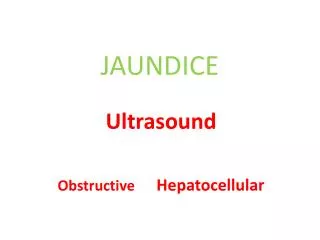
ABDOMINAL ULTRASOUND • Is a helpful diagnostic tool in the evaluation of neonatal cholestasis because it will identify • Choledocholithiasis • Perforation of the bile duct • Other structural abnormalities of the biliary tress such as a choledochal cyst
ABDOMINAL ULTRASOUND • In patients with biliary atresia, ultrasound may detect associated anomalies such as abdominal polysplenia and vascular malformations • The gallbladder is either not visualized or a microgallbladder is seen
HEPATOBILIARY SCINTIGRAPHY • Is used to differentiate biliary atresia from nonobstructive causes of cholestasis • The hepatic uptake of the agent is normal in patients with biliary atresia, but excretion into the intestine is absent • In neonatal hepatitis, though uptake may be impaired, excretion into the bowel will eventually occur • A follow-up scan after 24 hours is of value to determine the patency of the biliary tree
HEPATOBILIARY SCINTIGRAPHY • DRAWBACKS: • Though sensitive, it is not specific for biliary atresia • Fails to identify other structural abnormalities of the biliary tree or vascular anomalies