Download
1 / 35
350 likes | 457 Views
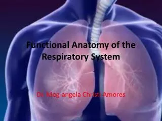
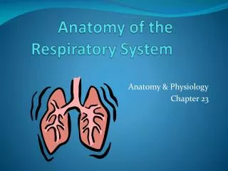
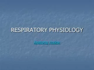
Respiratory Anatomy. Interesting Facts. The surface area of the lungs is about the same size as a tennis court You lose about ½ L of water a day through breathing Tuberculosis (TB) is caused by bacteria that destroy the air sacs in the lungs 2 million people die of TB each year.

E N D
Interesting Facts • The surface area of the lungs is about the same size as a tennis court • You lose about ½ L of water a day through breathing • Tuberculosis (TB) is caused by bacteria that destroy the air sacs in the lungs • 2 million people die of TB each year
Lizards can’t breathe when they are running . . . their breathing depends on the muscles between their ribs which MUST be used during running • The vapour that comes out of your mouth when you cough travels at about 160 km/h • Some animals (some frogs) can breathe through their skin
One acre of trees produces enough oxygen to keep 18 people alive for one year • Cigarettes and cigarette smoke contain over 4000 chemicals, including 43 known to cause cancer • Every cigarette shortens your life by ~14 minutes
The need for oxygen • Humans need oxygen to survive (250 mL/min). We can live several days without water, weeks without food, but only minutes without oxygen • Composition of atmosphere: 78% nitrogen, 21% oxygen, 0.03% carbon dioxide • Cells obtain energy by breaking down sugars; oxygen is needed for this to happen
The process of breaking down sugars into energy is called cellular respiration: C6H12O6 + 6O2 → 6H2O + 6CO2 + energy • The opposite is photosynthesis
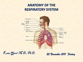
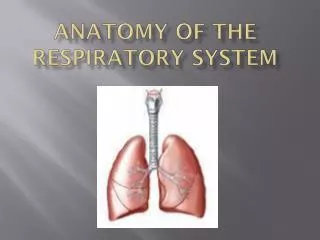
Nasal/oral cavity-> pharynx -> epiglottis -> larynx -> trachea -> bronchi ->bronchioles -> alveoli • Air enters through the nasal cavities or the mouth. Three important things happen: • Foreign particles are prevented from entering because of tiny hairs. • Air is warmed and moistened as it enters the body.
Nasal/oral cavity -> pharynx -> epiglottis -> larynx -> trachea -> bronchi -> bronchioles -> alveoli • From the nasal cavity, air travels through the pharynx (air filled channel in the mouth) into the larynx through the epiglottis. • Your tonsils are located in the pharynx
Nasal/oral cavity ->pharynx-> epiglottis -> larynx -> trachea -> bronchi -> bronchioles -> alveoli • The pharynx also opens into the esophagus where food travels to the stomach. • When food is chewed, it is forced to the top of the mouth, and pushed backwards. This forces the epiglottis to close, allowing food to enter the esophagus, not the trachea. • If you swallow too fast, cilia (hair-like protein structures) push particles out of respiratory tract and force a violent cough.
Nasal/oral cavity ->pharynx -> epiglottis -> larynx -> trachea -> bronchi -> bronchioles -> alveoli • Air travels through the larynx, commonly called the voice box. • Elastic ligaments create sound when air from the lungs is forced towards the pharynx. • The larynx is protected by a thick band of cartilage, commonly called the Adam’s Apple. The growth of this cartilage and larynx during puberty cause the deep voices of males.
Nasal/oral cavity ->pharynx -> epiglottis -> larynx -> trachea -> bronchi -> bronchioles -> alveoli • Air travels through the trachea (12 cm longs) and through right and left bronchi. These structures contain cartilaginous rings for support. • The bronchi lead to the right and left lung, leading air into the bronchioles.
Nasal/oral cavity ->pharynx -> epiglottis -> larynx -> trachea -> bronchi -> bronchioles -> alveoli • The bronchioles lead to the alveoli. • The alveoli are surrounded by capillaries. It is here where oxygen and carbon dioxide exchange takes place.
The Lungs • Well protected by the ribs, sternum and spine • Contained within the pleura, 2 membranous sacs which surround the lungs • The pleura help to isolate each lung
Smoking Commercials The Truth about Tabacco Please Quit Smoking Tabacco Wars Tabacco Wars-2 Tabacco Wars-3
For air to enter the lungs, 2 basic actions must occur: 1. The diaphragm – a thin, dome shaped sheet of muscle (~level with the bottom of the ribs), is curved upward in the middle, like an upside down saucer – as we breathe in, the sheet is pulled downward (flattens it out)
2. The second action causes the rib cage to move upward and outward – this results in contraction of the intercostals muscles which lie between the ribs
Inspiration (breathing in) • The volume of the lungs increases as the chest wall moves upward and outward, and the diaphragm moves downward • As the volume increases, pressure decreases; as the pressure decreases, air rushes in to equalize the pressure inside the lungs
The process of inspiration requires that muscles actively contract
Expiration (breathing out) • As the diaphragm relaxes, it pushes up to regain its shape • The intercostals muscles in the chest wall relax and the ribs move down and inward
These movements decrease the volume of the lungs, the pressure inside increases which pushes air out of the lungs until the internal and external pressure are equal once more • Breathing out requires no muscle contraction – it is just the result of muscle relaxation
Lung Capacity • Healthy adult – average 14-20 breathes per minute • The amount of air moved by a normal individual breathing while at rest is called the tidal volume – this is only a portion of the potential lung capacity
If you forcibly push out as much air as you can, the air you remove is called the expiratory reserve volume • Similarly the amount of extra air you can forcibly pull in is the inspiratory reserve volume
These three volumes together make up the vital capacity of the lungs • No matter how hard you try to push air out of the lungs, there will always be a small amount left in the spaces and tubes – called residual air capacity
Structure and Function • The structure and function of the respiration tract is to maximize air exchange, and minimize foreign particles from entering the lungs.
Repiratory system with the Circulatory System • Respiratory system brings oxygen into the body • Oxygen will cross the membranes, enter the bloodstream, and be transported to the cells which require oxygen for their activities