Download
1 / 28
460 likes | 1.05k Views
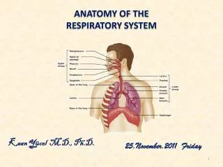
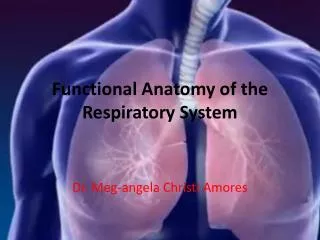
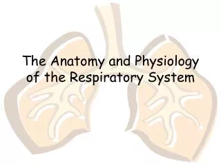
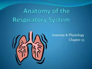
ANATOMY OF RESPIRATORY SYSTEM. DR SADIA FARHAN. Anatomy of the Respiratory System. Consists of all structures in the body that make up the airway and help us breathe Diaphragm Intercostal muscles Accessory muscles of breathing Nerves from the brain and spinal cord to those muscles.

E N D
ANATOMY OF RESPIRATORY SYSTEM DR SADIA FARHAN
Anatomy of the Respiratory System • Consists of all structures in the body that make up the airway and help us breathe • Diaphragm • Intercostal muscles • Accessory muscles of breathing • Nerves from the brain and spinal cord to those muscles
Anatomy of the Upper Airway • Upper airway • Airway structures above the vocal chords • Larynx • Divides upper and lower airways • Pharynx • Extends from the nose and mouth to the esophagus and trachea • Nasopharynx • Oropharynx • Laryngopharynx
Nasopharynx • Receives air from the nose • Formed by the union of facial bones • Lined with a ciliated mucous membrane • Keeps contaminants out of the respiratory tract • In illness, additional mucus traps agents
Nasopharynx • Divided into two passages by nasal septum • May be deviated • Numerous openings extend into the paranasal sinuses.
Nasopharynx • Paranasal sinuses • Prevent contaminants from entering respiratory tract • Fractures may cause: • Cerebrospinal rhinorrhea • Cerebrospinal otorrhea
Oropharynx • Forms the posterior of the oral cavity • Fracture or avulsion of teeth may result in aspiration • Tongue • Attached to mandible and hyoid bone • Most common cause of airway obstruction
Oropharynx • Palate • Separates oropharynx and nasopharynx • Hard palate and soft palate • Palatopharyngeal arch: entrance to the throat (pharynx)
Oropharynx • Tonsils • Adenoids and tonsils may become swollen and infected. • May cause upper airway obstruction
Glottis • Space between the vocal cords
Anatomy of the Lower Airway • Consists of the structures conducting the air from upper airway to alveoli • Exchanges oxygen and carbon dioxide
Larynx • Marks where the upper airway ends and lower airway begins • Thyroid cartilage • Formed by two plates that form the laryngeal prominence (Adam’s apple)
Larynx • Cricoid cartilage (cricoid ring) • First ring of the trachea • Cricothyroid membrane: ligament between the thyroid and cricoid cartilage • Site for emergency surgical and nonsurgical access to the airway (cricothyrotomy)
Glottis • Vallecula • Pocket between base of tongue and epiglottis • Important landmark for ET intubation • Arytenoid cartilages • Posterior attachment of the vocal cords • Valuable guides for ET intubation
Glottis • Piriform fossae • Airway devices are occasionally inadvertently inserted into these pockets • Laryngospasm: spasmodic closure of the vocal cords • Seals off the airway
Trachea • Conduit for air entry into the lungs • Begins below the cricoid cartilage • Descends down the midline of the neck and chest to the fifth or sixth thoracic vertebra • Esophagus lies posterior to the trachea • Trachea divides into right and left mainstem bronchi • Trachea and mainstem bronchi lined with: • Goblet cells • Cilia
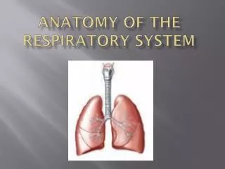
Lungs • Consist of the entire mass of tissue that includes smaller bronchi, bronchioles, and alveoli
Lungs • Hilum • it is the medial aspect of lung through which main bronchi and blood vessels enter the lung • Lobes of lungs • Right lung has three lobes and left lung has two lobes • Pleura • Lungs are covered with a thin lining which is called pleura. Pleura lining the lung is called visceral pleura and the outer layer is called parietal pleura which lines the chest wall
Pleural fluid • Small amount of fluid is present between the two layers of pleura which facilitates the lung movements • Bronchioles • Trachea divides into two bronchi, each of which enter the lung at hilum • Each main bronchus divides into branches called bronchioles • After 22 to 26 divisions the terminal branches of bronchus are called “terminal bronchioles”
Bronchioles contain smooth muscles and can constrict in response to certain stimului constriction of bronchioles is called “bronchoconstriction” • Alveoli • Functional site for the exchange of oxygen and carbon dioxide which increase surface area of the lungs • Lined with a phospholipid compound called surfactant. Surfactant deficiency can cause the alveoli to collapse ( atelectasis )