Download
1 / 67
710 likes | 801 Views
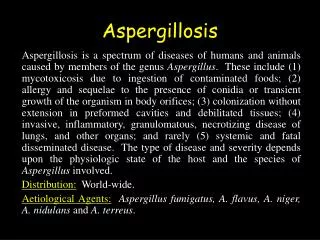
Aspergillosis. Dr Katherine Syred 14/07/14. Aspergillosis. Second most common human pathogen, generally infects the lungs via the airways Causes four major types of human disease. The different disease types affect different types of patient. invasiveness. What is Aspergillus?.

E N D
Aspergillosis Dr Katherine Syred 14/07/14
Aspergillosis • Second most common human pathogen, generally infects the lungs via the airways • Causes four major types of human disease. • The different disease types affect different types of patient
What is Aspergillus? • Fungi can exist in hyphae or spore form • Second most common fungal pathogen humans • Aspergillus is a mould and usually exists as hyphae in the body • It is a commensal in the environment and does not usually affect the healthy immunocompetent patient
Fungal Hyphae, 45-35˚acute angle branching, 7-10 microns diam.
In the lung parenchyma, little air is available to the fungi which grows anerobically by hyphae branching
Aspergillus has several sub-species including: • Aspergillus fumagatus, flavus, niger • Morphology- acute angle branching fungal hyphae, 35-45 degrees, 3-6um
Hypersensitivity Pneumonitis • “An immune mediated response to an extrinsic antigen which involves both immune complex and delayed hypersensitivity reaction” (Robbins and cottran)
Allergic Bronchopulmonary Aspergillosis (ABPA) • Specific large airways centred clinical syndraome, asthmatics 1-2% and CF (7-9% • Usually A. Fumigatus Immune mediated • History very important
How to diagnose allergic bronchopulmonary aspergillosis • Mechanism, production of cytokine IL 8 increased, can see eosinophils, T CD4/ CD8 • Bronchoalveolar lavage (BAL)– Cell count shows increase lymphocytes- • Th2 reaction (subset of CD4+) cells implicated • Proteolytic enzyme interrupts macrophage/ neutrophil killing hyphae/ spores
ABPA • Airway plugging and dilatation with eosinophils
Classical criteria diagnosis • Clinical deterioration • Immediate positive skin test • Ig E >1000kU/L • A. fumagatus + ppt • Altered CXR
What does this look like in the lung? • ABPA: Bronchi can show asthmatic changes (airway plugging/ smooth muscle hyperplasia) but often changes more pronounced in alveoli • Small numbers of hyphae in mucus • Numerous eosinophils/ cellular bedris in mucus
Gross specimen, airway plugging ABPA • Cylindrical bronchiectasis of central airways, become occluded by mucoid impaction. • Atelectasis of a lobe, often upper. Squamous metaplasia of airways.
Hypersensitivity Pneumonitis/ Extrinsic Allergic Alveolitis • General term for abnormal TH1 response in the lung to different allergens in different contexts (NOT just Aspergillus)
What is the clinical syndrome?1.Occupational high exposures may lead to alveolitis in otherwise normal individuals • The antigens are different in different environments: • Brewer’s lung (A Clavatus) • Farmers Lung (actinomyces) • Pigeon keepers lung (bird dander) • Popcorn workers lung (corn dust)
The spectrum allergic disease caused by Aspergillus (fumagatus) High exposure/ normal immunity Brewers lung occupational Hypersensitivity pneumonitis Aspergillus idiopathic Low exposure/ asthmatics/C.F Allergic bronchopulmonary aspergillosis
How is hypersensitivity pneumonitis diagnosed - 1 • HISTORY IS VITAL – detailed work and social history/ pets/ hobbies. May need inspection of home environment • HISTORY of the disease: • Exposure – Acute 4-6 hours, later fever/ SOB/ cough/ raised WCC • Alleviated by a spell in hospital
Diagnosis 2 • Bronchoalveolar lavage – Histology can do differential white cell count, raised is lymphocytes indicative and specific • Radiological appearance • Video-assisted thoracoscopic (VATS) biopsy if diagnosis is difficult
If the exposure to antigen continues, leads to progressive tissue damage, airway plugging, bronchocentric inflammation. • Later chronic changes characteristic multinucleate giant cells • Can lead to irreversible fibrosis (UIP like) • Vital to remove stimulating antigen
Early Hypersensitivity pneumonitis • Bronchocentric inflammation
Treatment • This is an abnormal/ excessive immune response to an allergen in the airways • Immunosupression will damped the bodies excessive T-Cell response to allergen • Steroids then cyclophosphmide etc • Will prevent progression • Also need to investigate house / work place for vital erradication of allergen source
Histological appearance Foreign body granulomata
If not treated adequately and stimulant antigen remains, ABPA/ Acute hypersensitivity pneumonitis can progress to Chronic hypersensitivity pneumonitis
Chronic hypersensitivty pneumonitis -leads to fibrosis If Chronic hypersensitivity pneumonitis not treated, progresses to end stage fibrosis “honeycomb lung” (asbestosis/ other fibrotic lung diseases can look identical)
Colonising Aspergilloma • Fungal Ball • Occurs in cavities previously formed by: -Old Tuberculosis -Abscess -Bronchiectasis • No real invasion of tissues • Different patients – previous cavitating disease
How do these patients present? • Recurrent haemopytysis • Hx can help • Very high aspergillus precipitin titre is vital Differential diagnosis of solitary pulmonary mass: ( get previous history) • Tuberculous mass • Carcinoma • Hamartoma (benign tumour)
Histology • Pink necrotic centre – dead cells and debris • Rim of active hyphae
Rim of hyphae Necrotic centre
Fruiting bodies • The air in the cavity allows the fungi to undergo aerobic reproduction
Fruiting Bodies Contain Spores • This is the ‘Aspergillium’
Treatment • Depends on severity of symptoms • Can be monitored over a number of years and observed if stable • May need FNAC/ biopsy to exclude other pathology • Can progress if patient become immunosupressed
Chronic Granulomatous Pneumonia • Chronic granulomatous infection of the lung by aspergillus species • A Bronchocentric granulomatous inflammation • Not angioinvasive, although can progress to this • Prominent formation of granulomata • (collections of macrophages)
By definition has not become invasive • Can progress to become invasive • Need for exposure history and early recognition/ treatment
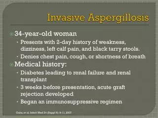
Invasive Aspergillosis • Usually originates from pulmonary infection either de novo or from chronic granulomatous aspergillosis • Lung is susceptible to these tiny spores in the environment
Who is susceptible • Haematology/ paediatric cancer units – especially if building work • The immunocompromised, especially neutropenia • Immunosupressed host – DM, Steroids, HIV, chemotherapy • Chronic illnesses – ie diabetes mellitus, asthma, mild immunosupression
How does Aspergillus colonise the body? • Breathed in through the airways • Grows in the moist airway into the interstitium of the lung • Angio-invasion is a key feature – vascular access leads to widespread dissemination
Patient’s CT scan (on methotrexate for psoriatic arthritis, CAPD for renal failure, also an amputee)
Can then spread to bowel wall, liver, kidneys • Devastating disease, difficult to treat with anti-fungals
How do we diagnose this? • Clinical history very important, low threshold for adding anti fungals immunosupression • High index of suspicion ie haematology/ oncology wards • In lung –imaging HRCT • Brochoalveolar lavage - BAL • Sputum • Biopsy – lung/ bowel etc
Laboratory tests • MICROBIOLOGY – sputum culture and probe tests • Serological tests – not yet in commercial use • (Can be difficult to detect) • Sputum cytology and bronchial biopsy histology – can be helpful • Communicate with your microbiologists!