Download
1 / 53
670 likes | 1.18k Views
ASPERGILLOSIS. A variety of diseases: pulmonary, external ears, eyes, meninges, sinuses or blood stream. ALLERGIC FUNGUS BALL INVASIVE. Clinical symptoms are not specific Radiography not specific (except fungus ball) Blood cultures seldom positive Serology seldom positive (early)

E N D
ASPERGILLOSIS A variety of diseases: pulmonary, external ears, eyes, meninges, sinuses or blood stream
ALLERGIC FUNGUS BALL INVASIVE
Clinical symptoms are not specific Radiography not specific (except fungus ball) Blood cultures seldom positive Serology seldom positive (early) Need invasive procedures for early detection Difficult to diagnose
A. FUMIGATUS A. NIGER A. FLAVUS
GEOGRAPHIC DISTRIBUTION WORLD-WIDE
SOIL DECAYING VEGETATION FOOD MEDICATION AIR VENTS DISINFECTANTS
More than 900 species Slow growing Various gross colors Spores Size Shape Texture color
DICHOTOMOUS BRANCHING WIDE, SEPTATE HYPHAE
Aspergilloma Cavity wall
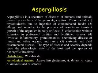
Aspergillosis is a spectrum of diseases of humans and animals caused by members of the genus Aspergillus. These include (1) mycotoxicosis due to ingestion of contaminated foods; (2) allergy to the presence of conidia or transient growth of the organism in body orifices; (3) colonization without extension in preformed cavities and debilitated tissues; (4) invasive, inflammatory, granulomatous, necrotizing disease of lungs, and other organs; and rarely (5) systemic and fatal disseminated disease. The type of disease and severity depends upon the physiologic state of the host and the species of Aspergillus involved. Distribution: World-wide. Aetiological Agents:Aspergillus fumigatus,A. flavus, A. niger, A. nidulans and A. terreus. Aspergillosis
Verycommon: compost heaps, airbone dustUsuallyaffects the lungs and sinusesLess often: very agressive, spread trough outthelungs and bloodstream to brain and kidneys
Aspergillosis–Symptoms& Diagnosis May cause no symptoms fungus ball Rx) Reapeted coughing up of blood (severebleeding) Fever Chestpain Difficulty breathing
Aspergillosis-Symptoms& Diagnosis Can affect deepertissues: 1.Renalfailure 2.Liverfailure 3.Shock 4.Delirium 5.Boodclots 6.Deathcanoccurquickly Chestx-ray Computedtomography(CT) Wheneverpossible : sendmaterialto a laboratoryto confirmidentification
Aspergilloma found at post-mortem in the lung of a child with leukaemia. Note fungus ball occupying cavity.
Aspergilloma found at post-mortem in the lung of a child with leukaemia.
Aspergilloma found at post-mortem in the lung of a child with leukaemia.
Aspergillosis in air sacs of a hen during an epidemic of aspergillosis in poultry.
Grocott’s methenamine silver (GMS) stained tissue section of lung showing fungal balls of hyphae of Aspergillus fumigatus.
Grocott’s methenamine silver (GMS) stained tissue section of lung showing dichotomously branched, septate hyphae of Aspergillus fumigatus.
Grocott’s methenamine silver (GMS) stained tissue sections showing Aspergillus fumigatus in lung tissue, note conidial heads forming in an alveolus.
Grocott’s methenamine silver (GMS) stained tissue sections showing Aspergillus fumigatus in lung tissue, note conidial heads forming in an alveolus.
Aspergillus fumigatus on Czapek dox agar showing typical blue-green surface pigmentation consisting of a dense felt of conidiophores.
038 Microscopic morphology of Aspergillus fumigatus showing typical columnar, uniseriate conidial heads. Conidiophores are short, smooth-walled and have conical shaped terminal vesicles, which support a single row of phialides on the upper two thirds of the vesicle. Conidia are produced in basipetal succession forming long chains (slide 038), however, during preparation of slides the conidial chains are usually disrupted giving the more typical microscopic appearance seen in slide 039. Conidia are globose to subglobose, green and rough-walled to echinulate
Microscopic morphology of Aspergillus fumigatus showing typical columnar, uniseriate conidial heads. Conidiophores are short, smooth-walled and have conical shaped terminal vesicles, which support a single row of phialides on the upper two thirds of the vesicle. Conidia are produced in basipetal succession forming long chains (slide 038), however, during preparation of slides the conidial chains are usually disrupted giving the more typical microscopic appearance seen in slide 039. Conidia are globose to subglobose, green and rough-walled to echinulate.
Aspergillus niger on Czapek dox agar. Colonies consist of a compact white or yellow basal felt covered by a dense layer of dark-brown to black conidial heads.
041 Microscopic morphology of Aspergillus niger showing large, globose, dark brown conidial heads, which become radiate, tending to split into several loose columns with age. Conidiophores are smooth-walled, hyaline or turning dark towards the vesicle. Conidial heads are biseriate with the phialides borne on brown, often septate metulae. Conidia are globose to subglobose, dark brown to black and rough-walled.
Aspergillus flavus on Czapek dox agar. Colonies are granular, flat, often with radial grooves, yellow at first but quickly becoming bright to dark yellow-green with age.
Microscopic morphology of Aspergillus flavus. Conidial heads are typically radiate, later splitting to form loose columns, biseriate but having some heads with phialides borne directly on the vesicle. Conidiophores are hyaline and coarsely roughened, often more noticeable near the vesicle. Conidia are globose to subglobose, pale green and conspicuously echinulate. Some strains produce brownish sclerotia.
Aspergillus nidulans on Czapek dox agar showing typical plain green colony with dark red-brown cleistothecia developing within and upon the conidial layer. Reverse may be olive to drab-grey or purple-brown.
Microscopic morphology of Aspergillus nidulans. Conidial heads are short columnar and biseriate. Conidiophores are usually short, brownish and smooth-walled. Conidia are globose and rough-walled.
Aspergillus terreus on Czapek dox agar showing typical suede-like cinnamon-buff to sand brown colonies. Reverse yellow to deep dirty brown.
Conidial head of Aspergillus terreus. Conidial heads are compact, columnar and biseriate. Conidiophores are hyaline and smooth-walled. Conidia are globose to ellipsoidal, hyaline to slightly yellow and smooth-walled.
Immunodiffusion test showing precipitins against Aspergillus.
Antifungal susceptibility disk test showing the in vitro activity of voriconazole against Aspergillus fumigatus with Candida krusei as a control.
Aspergillosis-Prognosis& Treatment Whenpresentin a sinus or single spot in the lung progresses SLOWLY Requires treatment, but no immediatedanger If infection widespread: patient isseriously ill Voriconazole Amphotericinb Itraconazole Capsofungin
If fungus balls grow near lungs .Blood vessels removed surgically Sinuses aspergillosis removed surgically Ear canal infection scrapingout the fungus and topics antifungal drops
Polyenes • ABLC, ABCD, • AmBisome • liposomal nystatin • inhaled amphotericin B • Azoles • itraconazole (i.v.) • voriconazole • posaconazole • ravuconazole • BAL 8557/4815 • Echinocandins • caspofungin • micafungin • anidulafungin • aminocandin Note: Blue text, earlier stage development Note: ABLC=amphotericin B lipid complex; ABCD= amphotericin B colloidal dispersion New (and newer) antifungals for invasive aspergillosis
Invasive Aspergillosis in Transplant Recipients Singh N & Paterson DL, Clin Microbiol Rev 2005;18:44-69.
918 Isolates; Jan. 2001-July 2004 AmB MFC >16 • A. fumigatus 24% AmB MIC>2 • A. terreus 90% • A. flavus 51% • A. ustus 50% A. nidulans 3% AmB=Amphotericin B; MFC=Minimum Fungicidal Concentration; MIC=Minimum Inhibitory Concentration Sutton D et al, Advances Against Aspergillus 2004 (Abstract 16) Aspergillus spp. Isolates Submitted to San Antonio Fungus Testing Laboratory
Polyene: broad spectrum of activity Lipid formulations of amphotericin B Reduced toxicities of intravenous amphotericin B deoxycholate Improved therapeutic index: ≥5 mg/kg/d well tolerated Salvage therapy (limited efficacy 40% responded) Empiric therapy (reduced efficacy vs moulds at lower doses) Limited data for primary therapy; most studies in empirical use Target use for patients with documented need (eg Zygomycosis, intolerance or progressive infection) Cost remains significant obstacle to use! Lipid Preparations of Amphotericin B: Rationale for Use
AmB Caspo Itra Amphotericin B0.15 g/mL Caspofungin0.30 g/mL Itraconazole2.6 g/mL ControlCells LivingCells Dead Cells In Vitro Effects of Echinocandins on Growth of Aspergillus In vitro activity: • Not classically fungicidal or fungistatic • Activity against other Aspergillus spp. (A terreus) • Animal models prolonged survival Bowman et al. Antimicrobial Agents Chemother 2002;46:3001-3012
Study conducted in patients with well-documented, refractory infection Efficacy Progressive infection Multiple prior antifungals Excellent tolerability Clinical utility for moulds Combination therapy (not primary therapy) Activity: Aspergillus (not Zygomycetes or other moulds) % % Echinocandins in Invasive Aspergillosis Maertens J et al. Clin Infect Dis 2004;39:1563-71
Consideration of risk Role of mycological diagnosis Predictive value of positive cultures in high risk patients Utility of radiological procedures Non-culture based diagnostics Impact of antifungal therapies Value of serial samples Significance of false negative/false positive results Role of testing in other body fluids, including CSF & BAL Role of surrogate markers in decision making & impact on mortality Diagnostic Strategies in Invasive Aspergillosis
In vitro Most interactions show synergy / additive effects (Perea, 2002) Poor correlation between in vitro results and in vitro efficacy (Johnson, 2004) Experimental infections Candin plus polyene (Kohno, 2000; Nakajima, 2000) Candin plus azole (Kirkpatrick, 2002; Petraitiene, 2002) Improved sterilization of tissues Reduced tissue burden Anecdotal clinical series Candin+polyene (Aliff, 2003; Kontoyiannis, 2003; Ratanatharathorn, 2002) Candid plus azole (Marr, 2004) Combination Therapy: Candins