Download
1 / 28

E N D
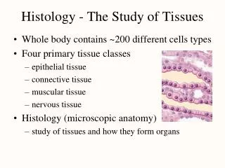
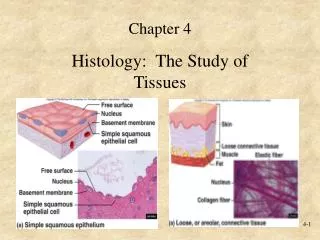
Histology:**is the study of tissue sectioned as a thin slice, using a microtome.**Tissue is taken from a french tissu (weave or texture), referring to a cloth or fabric, which in turn meaning tissu derived from the Greek istos, meaning tissue or web.**and "-logos" = the study of.**Histology can be described as microscopic anatomy.**Histology is an essential tool of biology.
The Science Of Histology Deals With: • the microscopic and ultramicroscopic anatomy of the fundamental cells, tissue, and organs of the body, • and in its broader aspect, correlates structural features with function.
Microscope and basic histology • The light microscopeis still the most common type of microscope used in basic histology • The phase contrast microscope,used especially for living cells; • The dark field microscopewhich uses a special condenser for producing a dark field to enhance contrast; • Microscopes that utilize ultraviolet rays, x-rays, and polarized lightfor observing specimens and enhancing resolution, density, or birefringence. • Electron microscope: *TEM two dimensional image & *SEM three dimensional image reveals surface contours of the specimen
Introduction to cells and tissues • The vital functional biological units of living matter are the cells (a term coined by Robert Hooke, 1665). • All living things are composed of cells and cell products.
The Animal Cell Is the typical eukaryotic cell
Animal Cell Structure • enclosed by a plasma membrane and • containing a membrane-bound nucleus and organelles. • Unlike the eukaryotic cells of plants and fungi, animal cells do not have a cell wall. • Most cells, both animal and plant, range in size between 1 and 100 micrometers and are thus visible only with the aid of a microscope.
Struture of the nucleus • Nuclear envelope • Karyosol • Chromatincomposed of: Nucleoproteins and DNA plus RNA *Euchromatin is the long thin chromatin threads form a network of diffuse thin nucleoprotein threads which represent active regions on DNA strands where transcription can take place. *Heterochromatin is the condensed regions represent coiled chromatin and is inactive DNA. heterochromatin is markedly stained by basic dyes and is increasingly evident near the end of the cell’s life Nucleolus
Animal Cell Nucleus 1- present or absent • present: in all animal cells except, • abcent: in mature human erythrocytes Cells lacking a nucleus cannot undergo cell division, and as a result , have a limited lifespan.
2- Nuclear shape • Sperical, Lobulated, • Ovoid, Segmented • Indented, Elongated 3- Nuclear size: Normally range from 3 to 14µm in diameter
Peripheral blood smear of toad The red blood cells are nucleatied
Human blood smear:Most all the cells you see in this picture are red blood cells (erythrocytes), and the blue and green arrows are pointing to white blood cells (leukocytes). • Erytheocytes are unnucleated • Leukocytes are nucleated cells
The nuclei of the white blood cells manifest different shapes.
4- Number of nucleus: • Mononucleated (most cells) • Binucleated (cardiac cells or parenchymal liver cells) • Multinucleated (osteoclast and skeletal muscle cells)
5- Location of the nucleus • Central • Eccentric • Peripheral • Basal
Skeletal Muscle - multinucleatedcylinder like cellThe striations (stripes) are due to a very orginized pattern of proteins found within the cells. The black arrow is pointing to a nucleus.
Animal Cell Structure • Cells of multicellular organisms, in meeting the demands of specialization of labor, have evolved many structural features to accommodate various functional needs, for examples:
I- Egg cell :are relatively large in order to accommodate supplies of yolk. The mammalian oocyte is held in a quiescent state in the surrounding follicle until it begins to grow during the ovulatory cycle.
II- Sperm cells:are small since they are composed mostly of nuclear material and motile; Human sperm cells stained with Hematoxylin and Eosin (H&E)
III- Nerve cells:are elongate because of their axons which are needed to connect other neural components; Diagram of nerve cell
IV- Fat cells • are usually large because of stored lipids.
V-Smooth muscle cells (fibers) are spindle shaped cells that form masses.
VI-Cardiac muscle fibers - or heart muscleFound only in the heart! Like skeletal muscle, cardiac muscle is striated, but cardiac muscle also contains structures called intercalated disks (blue arrow).
Hepatocyte nucleus sinusoid Central vein sinusoid sinusoid Endothelial cell nucleus VII- Liver, cuboidal cells
Kupffer cells Space of Disse (perisinusoidal space) hepatocyte liver; sinusoids and Kupffer cells