Download
1 / 17
410 likes | 1.2k Views
Transmission Electron Microscope. TEM Image of E. Coli. TEM Image of Mitochondria. TEM Image of Computer Chip. SEM Image compared to TEM Image. Transmission Electron Microscope. http://www.matter.org.uk/tem/ http://em-outreach.ucsd.edu/web-course/toc.html

E N D
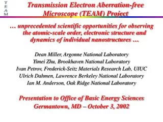
Transmission Electron Microscope FNI 2C EM
TEM Image of E. Coli FNI 2C EM
TEM Image of Mitochondria FNI 2C EM
TEM Image of Computer Chip FNI 2C EM
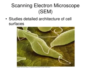
SEM Image compared to TEM Image FNI 2C EM
Transmission Electron Microscope • http://www.matter.org.uk/tem/ • http://em-outreach.ucsd.edu/web-course/toc.html • http://www.amazon.com/Transmission-Electron-Microscopy-Textbook-Materials/dp/038776500X/ref=sr_1_1?ie=UTF8&s=books&qid=1224191760&sr=8-1 FNI 2C EM
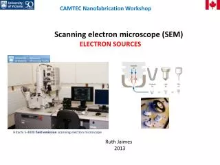
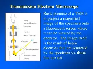
TEM Operation FNI 2C EM
UW Eau Claire’s TEM FNI 2C EM
UW Eau Claire’s TEM FNI 2C EM
TEM Phosphor Screen FNI 2C EM
TEM Control Panel FNI 2C EM
TEM Grids All TEM’s ever done have examined only .3 mm3 of material FNI 2C EM
TEM Components FNI 2C EM
TEM Sample Preparation and Theory of Operation • Samples for TEM must be electron transparent. That means they must be thin enough for electrons to pass through. • It is very difficult to achieve this. • After the sample has been polished very thin an ion mill is used to perform the final stages. • An ion mill uses an argon plasma to remove material from the sample. • High energy argon ions strike the sample and atoms of the sample are knocked away. • TEMs can be used to image atoms. • Some scientists have combined TEM chambers with reactors so they can watch chemical reactions in progress. FNI 2C EM
FIB Sample Preparation for TEM FNI 2C EM
Electron Microscope Vendors • JEOL http://www.jeol.com/ • Hitachi http://www.hitachi-hitec-uk.com/ • Zeiss www.smt.zeiss.com/nts • FEI www.feicompany.com • Camscan www.camscan-usa.com • Elektronen-Optik www.eos-do.com • Tescan www.tescan.com • KLA/Tencor/Amray http://www.kla-tencor.com/ FNI 2C EM
Notes • Growth of carbon nanotubes inside environmental TEM. FNI 2C EM