Download
1 / 9
170 likes | 470 Views
Electron Microscope. The resolution of a microscope is limited by the diffraction of light. Single diffraction The minimum size observable with visible light is about 200 nm. Airy disk Visible light down to 400 nm. Light Resolution. Small wavelength – no diffraction.

E N D
The resolution of a microscope is limited by the diffraction of light. Single diffraction The minimum size observable with visible light is about 200 nm. Airy disk Visible light down to 400 nm Light Resolution Small wavelength – no diffraction Large wavelength – diffraction
The diffraction limit of an electron is based on its wavelength. Related to momentum Energy of 110 V creates a wavelength of 0.117 nm. Nonrelativistic energy Rest energy 0.511 MeV hc = 1240 eV nm Electron Diffraction
Microscope Revisited • An optical microscope uses light to form an image. • The objective and eyepiece are the primary lenses. • A condenser lens is sometimes used to focus light onto the object. eyepiece intermediate image objective
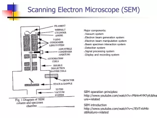
Electron Gun • A hot filament produces electrons attracted to the positive anode. • The Whenelt cap uses a negative potential to focus electrons onto the axis. • Potential about 500 V • Space charge electrons from 1 mm gap towards anode • Electrons accelerate through a gap in the anode plate. • Point source beam for optics • Monochromatic beam Images: University of Nebraska, Lincoln
Electrons are charges particles. Bent by magnetic fields Circular magnets act as lenses. Spherical lens equations Apertures are thin metal disks to block scattered electrons. Typical hole size 2-100 mm Electron Lens object image objective aperture intermediate image projector magnified image
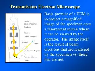
Transmission • A transmission electron microscope passes electrons through a sample. • The source is an electron gun. • Condenser lenses and create a narrow coherent beam. • Spot size lens • Intensity lens • Adjustable aperture • Intermediate and projector lenses increase magnification. • Image recorded on screen. • Light areas - more electrons Images: University of Nebraska, Lincoln
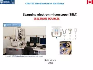
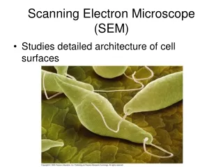
Scanning • A scanning electron microscope reflects electrons off of a sample. • The electron gun and condenser create the electron beam. • Fine and course current control • Electric coils sweep the beam over the sample. • Microsecond time scale • The objective lens targets a spot on the sample. • Reflection measured Images: University of Nebraska, Lincoln
Electron microscopes are able to make images with many times the detail of optical microscopes. At right, a mosquito head with an SEM. Top at x200 magnification Bottom at x1000 magnification Images next Museum of Science, Boston