Download
1 / 39
390 likes | 501 Views
Chapter 4: The Central Nervous System Part 3 – Brain research methods. Unit 3 – Area of Study 1 Mind, brain and body Pages 132-176. Study Design Content.

E N D
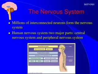
Chapter 4: The Central Nervous SystemPart 3 – Brain research methods Unit 3 – Area of Study 1 Mind, brain and body Pages 132-176
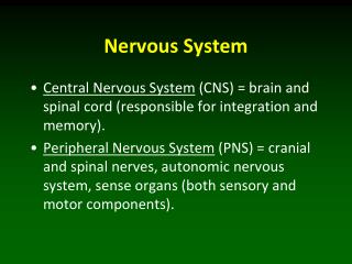
Study Design Content • the application and use of brain research methods in investigating the relationship between biological and cognitive factors of human behaviours including: – direct brain stimulation and transcranial magnetic stimulation (TMS) – brain recording and imaging techniques: computed tomography (CT), positron emission tomography (PET), single photon emission computed tomography (SPECT), magnetic resonance imaging (MRI), and functional magnetic resonance imaging (fMRI) • research methods and ethical principles associated with the study of the brain and states of consciousness, as outlined in the introduction to the unit
Brain Research Methods • Manual Activity 18 – Surfin’ the net – Finding out about brain research methods, pg. 31 • A major issue which presented problems for early researchers who were interesting in studying the function of the brain was the lack of technology • Early research was focused on the structure of the brain using brains donated from deceased individuals and animals • New technologies in the 20th century have provided researchers with an advanced understanding of how the brain functions
Brain Research Methods • Direct brain stimulation • Electrode stimulation • Transcranial Magnetic Stimulation (TMS) • Electroencephalograph (EEG) • Computerised Tomography (CT) • Magnetic Resonance Imaging (MRI) • Positron Emission Tomography (PET) • Single Photon Emission Computed Tomography (SPECT) • Functional Magnetic Resonance Imaging (fMRI)
Direct Brain Stimulation • Direct brain stimulation involves using a device that emits a weak electric current to activate or disrupt the normal activity of neurons in a specific brain area • We will examine two types of brain stimulation: • Electrode stimulation • Transcranial magnetic stimulation (TMR)
Electrode Stimulation • In the 1940’s, Wilder Penfield began to explore areas of the cerebral cortex associated with sensations and memory. • Penfield was able to ‘map’ areas of the cerebrum responsible for specific functions. • When a patient underwent open brain surgery, Penfield with permission attached small tags to particular parts of the brain thought to be responsible with certain functions.
Electrode Stimulation • Penfield then electrically stimulated each area and noted the responses made by his awake and alert patients. • During this process particular involuntary movements were made or speech was interrupted. This allowed Penfield to conclude that certain parts of the brain were responsible for certain functions. • Fig 4.55 pg.239 • Fig 4.56 pg.240 • Fig 4.57 pg.241
Value & Limitations of Electrode Stimulation • Values • Electrode stimulation has proved to be an effective technique in that it has enabled researchers to identify the locations and functions of numerous brain structures and areas, as well as hemispheric specialisation for different functions. • Limitations • One limitation of ESB is that it can only be completed by people undergoing brain surgery and that the process is extremely invasive. • No healthy control • Also the process imposes risks that today’s ethical standards would consider unacceptable.
Transcranial Magnetic Stimulation (TMS) • Transcranial magnetic stimulation (TMS) is a general term for a direct brain-stimulation technique that delivers a magnetic pulse through the skull and temporarily activates or disrupts the normal activity of neurons in a specific area of the cerebral cortex • While receiving the stimulation the participant is awake and alert as TMS is a non-invasive procedure and no anaesthetics or substances need be administered • When TMS is used in procedures involving the delivery of a single pulse it is called single pulse TMS or non-repetitive TMS – this is in contrast to repetitive TMS or rTMS which is used in procedures involving repeated delivery of a pulse
Transcranial Magnetic Stimulation (TMS) • Researchers use TMS to study functions of specific areas of the cerebral cortex • The pulse only affects the part of the brain which lies directly beneath the skull (2-3cm)
Values and Limitations of TMS • Values • A non-invasive procedure – the patient is completely alert and awake • Free of side-effects • Able to influence many brain functions • Limitations • As it induces an electrical current in the brain. TMS can cause a seizure although this risk is very low • There is slight discomfort coming from the stimulation of the scalp • Poor localisation – does not reach far into the brain, only the cortex
EEG - Electroencephalograph • An electroencephalograph (EEG) is a device that detects, amplifies and records general patterns of electrical activity of the brain. • The electrical activity is recorded by small electrodes attached to the scalp. • These electrodes detect any activity and graphs it on a computer. • The brain wave patterns recorded by the computer are called rhythms and are assigned the Greek letters – alpha, beta, delta, theta.
EEG - Electroencephalograph • The EEG has been widely used to detect various brain related medical conditions including: • Brain damage and neurological disorders. • Epilepsy • Parkinson’s Disease • Depression • Schizophrenia • Apraxia
Value and Limitations of the EEG • Values • The EEG is useful in providing information about brain wave activity without being invasive. • It is relatively easy and cost effective to use an EEG. • It can also be used for lengthy periods of time and by people of all ages. • Non-invasive procedure • Learning Activity 4.24 – Review Questions, pg. 248 • Limitations • Electrical activity can actually be lessened as the message must travel through the scalp, skin and headpiece. • Conducting the test on small children can be an issue as they may be afraid of the equipment, and they are also required to sit reasonably still. • Can at times be difficult to specifically pinpoint where brain wave activity is occurring. • Not an imaging technique
Computerised Tomography (CT) • Computerised Tomography (CT)is a neuro-imaging technique that produces a computer enhanced image of a cross section (slice) of the brain from x-rays taken from different angles. • The procedure involves moving an x-ray source in an arc around the head while a computer compiles different images of the brain and area investigated.
Computerised Tomography (CT) • The patient must be given an injection first which places an iodine contrast into the bloodstream. • This substance enables the major blood vessels in the brain to be highlighted. It is not radioactive and does not cause any side effects. • CT scans aid psychologists by detecting and showing things such as: • Strokes, tumors, injuries and other brain disorders. • Abnormalities associated with depression, Alzheimer’s, Schizophrenia and other disorders.
Values and Limitations of CT • Values • Non-invasive procedure • Provides detailed structural images of the brain • Research used on healthy and clinical participants • Abnormalities can be located • Limitations • Only provides horizontal pictures • Only shows structure not function • Expensive and needs trained staff to use
Magnetic Resonance Imaging (MRI) • Magnetic Resonance Imaging (MRI) is a neuroimaging technique that uses harmless magnetic fields and radio waves to vibrate atoms in the brain’s neurons to produce an image of the brain. • These vibrations are detected by a huge magnet in the chamber surrounding the person, and are channeled into a computer. • MRI is more sensitive than CT and therefore provides a clearer and more detailed image.
Magnetic Resonance Imaging (MRI) • MRI has primarily been used for diagnosing structural abnormalities of the brain. • It also has been used to detect small changes in the brain – for example detecting cancerous and non-cancerous cells. • MRI is also useful in the detection and diagnosis of degenerative diseases in the brain and central nervous system.
Values and Limitations of MRI • Values • The development of MRI technology in the last decade has enabled more precision in the study of the brain, that is accurate and non-invasive. • 3D imaging • Used in healthy and clinical settings • Learning Activity 4.26 – Review questions, pg.252 • Limitations • One limitation of MRI is that it cannot be used on patients that have internal metallic devices (pacemakers – bone pins). • Also like CT, MRI can only show brain structures – not function. • Very expensive
Positron Emission Tomography (PET) • Positron Emission Tomography (PET) produces a computer generated image that provides information about brain function and activity during various tasks. • PET is used to measure levels of activity in different areas of the brain while the participant is involved in a cognitive or behavioural activity of some kind, such as thinking, imagining, remembering, talking or moving a body part.
Positron Emission Tomography (PET) • Prior to the procedure a harmless radioactive substance is injected into the patients blood vessels. • This substance emits tiny radioactive messages which is detected by a sensitive electronic device connected to the computer. • Each PET scan uses a colour code to detect levels of activity. • Activity levels from lowest to highest are recorded in the following colours – violet, blue, green, yellow and red.
Values & Limitations of PET • Values • The major advantage of PET is that it offers detailed images of the functioning brain. • It can also be used on people with intact brains rather than just people with damaged brains. • Sensitive – can potentially image the entire brain, deep and shallow structures • Limitations • Even though the risks from the radioactive dye is minimal, exposure to PET must be kept short. • Researchers must also be aware that changes in brain activity may not always be due to the tasks set for the patient. • Very expensive • Mildly invasive due to the injection of the dye
Single Photon Emission Computed Tomography (SPECT) • A variation of PET, single photon emission computated tomography (SPECT) uses a longer lasting radioactive tracer and a scanner to record data that a computer uses to construct a 2D or 3D image of the brain • Similar to PET, SPECT images are functional in nature • SPECT images are not as good as PET images but are able to be combined with CT images to create a SPECT-CT image when a higher resolution is required or when a structure is needed to be located on the SPECT image
Values and Limitations of SPECT • Values • Provides and image of brain activity and function • Used for healthy and clinical patients • Less expensive than PET • Longer lasting radioactive isotope than PET • Limitations • Mildly invasive due to the injection of dye • The use of radioactive agents means only a limited number of scans can be performed on a subject • Less sensitive than PET
Functional Magnetic Resonance Imaging (fMRI) • fMRI is a neuroimaging technique that enables the identification of brain areas that are particularly active during a given task by detecting changes in oxygen levels in the blood flowing through the brain. • When an area of the brain is active, there is an increased oxygen flow to that area. • fMRI scans can be manipulated by a computer to also make 3D images of the functioning brain.
Values and Limitations of fMRI • Values • Unlike PET, fMRI doesn’t require any exposure to radiation. The images obtained also by fMRI are also more detailed than PET. • fMRI can also be used on patients with intact or damaged brains. • The time needed to take the images is also a lot faster which enables more images in a smaller time frame. This in turn allows more information gained about a specific brain function. • Limitations • Cannot map neuroreceptors as PET due as it is based on blood flow • Technique can be very expensive • Not subject friendlly
Brain Imaging Techniques • Learning Activity 4.27– Review questions, pg.259 • Learning Activity 4.28– Summarisingbrain research methods, pg. 259
Ethical Principles in Brain Research • As with any psychological study the following guidelines of participant’s rights must be adhered to: • Confidentiality • Voluntary Participation • Withdrawal Rights • Informed Consent Procedures • Deception • Debriefing
Ethical Principles in Brain Research • Also stated in the ‘National Statement on Ethical Conduct in Research Involving Humans’ are the requirements: • Integrity • Respect for Persons • Beneficence • Justice
Ethical Principles in Brain Research • Clearly, brain research raises many important ethical questions and issues, particularly the medical procedures that are often involved. • Researchers must ensure all ethical principles and guidelines promoted by the safeguarding policies are followed. • As a secondary precaution, all brain research studies must be approved by an Ethics Committee to allow the research to take place.
Ethical Principles in Brain Research • Box 4.18– Lobotomy, pg. 262 • Learning Activity 4.31– Applying Ethical Principles in Brain Research, pg. 263 • Chapter 4 Test, pg. 265