Download
1 / 14
160 likes | 981 Views
Polycythemia and Hyperviscosity. Kirsten E. Crowley, MD June, 2005. Definitions. Polycythemia is increased total RBC mass Central venous hematocrit > 65% Above 65% blood viscosity rises exponentially

E N D
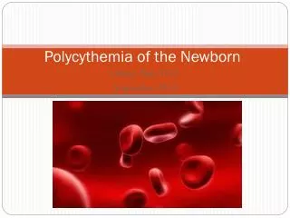
Polycythemia and Hyperviscosity Kirsten E. Crowley, MD June, 2005
Definitions • Polycythemia is increased total RBC mass • Central venous hematocrit > 65% • Above 65% blood viscosity rises exponentially • Polycythemic hyperviscosity is increased viscosity of the blood resulting from increased numbers of RBCs • Not all polycythemic infants have symptoms of hyperviscosity
Incidence • Polycythemia occurs in 2-4% of newborns • Half of these are symptomatic • Hyperviscosity occurs in 25% of infants with hematocrit 60-64% • Hyperviscosity without polycythmia occurs in 1% (nonpolycythemic hyperviscosity)
Pathophysiology • Clinical signs result from regional effects of hyperviscosity and from the formation of microthrombi • Tissue hypoxia • Acidosis • Hypoglycemia • Organs affected: CNS, kidneys, adrenals, cardiopulmonary system, GI tract
What affects hyperviscosity? • Hematocrit • Increased hct is the most important single factor • Results from increase in circulating RBCs or decreased plasma volume (dehydration) • Plasma viscosity • Higher plasma proteins = increased viscosity • Especially fibrinogen (typically low in neonates) • Not usually an issue in neonates • RBC aggregation • Occurs in areas of low blood flow = venous microcirculation • Not a large factor in neonates • Deformability of RBC membrane: usually normal
Conditions that alter incidence • Altitude: increased RBC mass • Neonatal age • Physiologic increase in hematocrit due to fluid shifts away from intravascular compartment with maximum at 2-4 hours of age • Obstetric factors: delayed cord clamping or “stripping” of the umbilical cord • High-risk delivery, especially if precipitous
Perinatal processes • Enhanced fetal erythropoiesis usually related to fetal hypoxia • Placental insufficiency • Maternal hypertension, abruption, post-dates, IUGR, maternal smoking • Endocrine disorders: due to increased oxygen consumption • IDM (>40% incidence), congenital thyrotoxicosis, CAH, Beckwith-Wiedemann syndrome (hyperinsulinism)
Hypertransfusion • Delayed cord clamping • Placental vessels contain 1/3 of the fetal blood volume, half of which will be returned within 1 minute • Gravity: positioning below the placenta will increase placental transfusion • Meds: oxytocin can increase contractions and thus transfusion • Decreased in c-section b/c no contractions • Twin-twin transfusion • Maternal-fetal transfusion • Intrapartum asphyxia • Enhances net umbilical flow toward the infant, while acidosis increases capillary leak leading to reduced plasma volume
Clinical presentation • Symptoms are non-specific! • CNS: lethargy, hyperirritability, proximal muscle hypotonia, vasomotor instability, vomiting, seizures, cerebral infarction (rare) • Cardiopulmonary: respiratory distress, tachycardia, CHF, pulmonary hypertension • GI: feeding intolerance, sometimes NEC • GU: oliguria, ARF, renal vein thrombosis, priapism • Metabolic: hypo-glycemia/-calcemia/-magnesemia • Heme: hyperbili, thrombocytopenia • Skin: ruddiness
Diagnosis • Central venous hematocrit > 65% • ALWAYS draw a central venous sample if the capillary hematocrit is > 65% • Warmed capillary hematrocrit > 65% only suggestive of polycythemia
Management • Asymptomatic infants • Expectant observation unless central venous hematocrit >75% (consider partial exchange transfusion) • Can do a trial of rehydration over 6-8 hr if dehydrated • Usually at > 48 hours of age and weight loss > 8-10% • Give 130-150 ml/kg/d • Check central hematocrit q6 hours • Normal peak is at 2-4 hours of age for acute polycythemia
Management • Symptomatic infants with central hct > 65% • Partial exchange transfusion is advisable but debatable • For exchange can use normal saline, Plasmanate, 5% albumin, or FFP • Volume exchanged = • (Weight (kg) x blood volume) x (hct - desired hct) / hct • Blood volume is 80 ml/kg • Exchange can be done via UVC that is not in the liver, low UAC, or PIV
Other labs to check • Serum glucose • Hypoglycemia is common with polycythemia • Serum bilirubin • Increased bili due to increased RBC turnover • Serum sodium, BUN, urine specific gravity • Usually high if baby is deyhdrated • Blood gas to rule-out inadequate oxygenation as cause of symptoms • Platelets, as thyrombocytopenia can be present • Serum calcium b/c hypocalcemia can be seen
Prognosis • Increased risk of GI disorders and NEC with partial exchange transfusion (PET) • Older trials show decreased neurologic complications from hyperviscosity with PET, but newer trials show no real benefit • PET is controversial! • Infants with asymptomatic polycythemia have an increased risk for neurologic sequelae • Normocythemic controls with the same perinatal history have a similarly increased risk