Download
1 / 43
440 likes | 645 Views
Circulatory Systems (Ch. 42). Take a look at a skeleton and see how well a heart is protected — open heart surgery takes breaking a body to get to the heart. Exchange of materials. Animal cells exchange material across their cell membrane fuels for energy nutrients oxygen

E N D
Circulatory Systems (Ch. 42) Take a look at a skeleton and see how well a heart is protected — open heart surgery takes breaking a body to get to the heart.
Exchange of materials • Animal cells exchange material across their cell membrane • fuels for energy • nutrients • oxygen • waste (urea, CO2) • If you are a 1-cell organism that’s easy! • diffusion • If you are many-celled that’s harder
Overcoming limitations of diffusion CO2 CO2 O2 NH3 aa NH3 CO2 NH3 CO2 CO2 NH3 O2 NH3 CO2 CO2 CO2 aa NH3 NH3 NH3 CHO CO2 CO2 aa CH • Diffusion is not adequate for moving material across more than 1-cell barrier aa O2 CH CHO CO2 aa NH3 CHO CH O2 aa
In circulation… • What needs to be transported • nutrients & fuels • from digestive system • respiratory gases • O2 & CO2 from & to gas exchange systems • intracellular waste • waste products from cells: water, salts, nitrogenous wastes • protective agents • immune defenses • regulatory molecules • hormones
Circulatory systems • All animals have: • circulatory fluid = “blood” • tubes = blood vessels • muscular pump = heart open closed hemolymph blood
Open circulatory system • Taxonomy • invertebrates • insects, arthropods, mollusks • Structure • no separation between blood & interstitial fluid • hemolymph
The fact that open and closed circulatory systems are each widespread among animals suggests that both offer advantages. For example, the lower hydrostatic pressures associated with open circulatory systems make them less costly than closed systems in terms of energy expenditure. Furthermore, because they lack an extensive system of blood vessels, open systems require less energy to build and maintain. And in some invertebrates, open circulatory systems serve a variety of other functions. For example, in molluscs and freshly molted aquatic arthropods, the open circulatory system functions as a hydrostatic skeleton in supporting the body.
Closed circulatory system • Taxonomy • invertebrates • earthworms, squid, octopuses • vertebrates • Structure • blood confined to vessels & separate from interstitial fluid • 1 or more hearts • large vessels to smaller vessels • material diffuses between blood vessels & interstitial fluid closed system = higher pressures
What advantages might be associated with closed circulatory systems? Closed systems, with their higher blood pressure, are more effective at transporting circulatory fluids to meet the high metabolic demands of the tissues and cells of larger and more active animals. For instance, among the molluscs, only the large and active squids and octopuses have closed circulatory systems. And although all arthropods have open circulatory systems, the larger crustaceans, such as the lobsters and crabs, have a more developed system of arteries and veins as well as an accessory pumping organ that helps maintain blood pressure. Closed circulatory systems are most highly developed in the vertebrates.
Vertebrate circulatory system • Adaptations in closed system • number of heart chambers differs 2 3 4 high pressure & high O2to body low pressureto body low O2to body What’s the adaptive value of a 4 chamber heart? 4 chamber heart is double pump = separates oxygen-rich & oxygen-poor blood; maintains high pressure
Evolution of vertebrate circulatory system Gill capillaries Lung and skin capillaries Lung capillaries Lung capillaries AMPHIBIANS REPTILES (EXCEPT BIRDS) MAMMALS AND BIRDS FISHES Right systemicaorta Pulmonarycircuit Artery Pulmocutaneouscircuit Pulmonarycircuit Gillcirculation Heart:ventricle (V) Left Systemicaorta A A A A A A Atrium (A) V V V V V Left Right Left Left Right Right Systemiccirculation Systemic circuit Systemic circuit Vein Systemic capillaries Systemic capillaries Systemic capillaries Systemic capillaries Birds ANDmammals! Wassssup?!
Evolution of 4-chambered heart • Selective forces • increase body size • protection from predation • bigger body = bigger stomach • endothermy • can colonize more habitats • flight • decrease predation & increase hunting • Effect of higher metabolic rate • greater need for energy, fuels, O2, waste removal • endothermic animals need 10x energy • need to deliver 10x fuel & O2 to cells convergentevolution
Vertebrate cardiovascular system • Chambered heart • atrium = receive blood • ventricle = pump blood out • Blood vessels • arteries = carry blood away from heart • arterioles • veins = return blood to heart • venules • capillaries = thin wall, exchange / diffusion • capillary beds = networks of capillaries
Arteries, veins, and capillaries are the three main kinds of blood vessels, which in the human body have a total length of about 100,000 km. • Notice that arteries and veins are distinguished by the direction in which they carry blood, not by the characteristics of the blood they contain. All arteries carry blood from the heart toward capillaries, and veins return blood to the heart from capillaries. A significant exception is the hepatic portal vein that carries blood from capillary beds in the digestive system to capillary beds in the liver. Blood flowing from the liver passes into the hepatic vein, which conducts blood to the heart.
Blood vessels arteries veins artery arterioles venules arterioles capillaries venules veins
Arteries: Built for high pressure pump • Arteries • thicker walls • provide strength for high pressure pumping of blood • narrower diameter • elasticity • elastic recoil helps maintain blood pressure even when heart relaxes
Veins: Built for low pressure flow • Veins • thinner-walled • wider diameter • blood travels back to heart at low velocity & pressure • lower pressure • distant from heart • blood must flow by skeletal muscle contractions when we move • squeeze blood through veins • valves • in larger veins one-way valvesallow blood to flow only toward heart Blood flows toward heart Openvalve Closed valve
Capillaries: Built for exchange Capillaries Venule Arteriole (a) Sphincters relaxed Arteriole Venule (b) Sphincters contracted Thoroughfare channel Precapillary sphincters • Capillaries • very thin walls • lack 2 outer wall layers • only endothelium • enhances exchange across capillary • diffusion • exchange between blood & cells (c) Capillaries and larger vessels (SEM) 20 m
Controlling blood flow to tissues • Blood flow in capillaries controlled by pre-capillary sphincters • supply varies as blood is needed • after a meal, blood supply to digestive tract increases • during strenuous exercise, blood is diverted from digestive tract to skeletal muscles • capillaries in brain, heart, kidneys & liver usually filled to capacity Why? sphincters open sphincters closed
Exchange across capillary walls Fluid & solutes flows out of capillaries to tissues due to blood pressure • “bulk flow” Lymphatic capillary • Interstitial fluid flows back into capillaries due to osmosis • plasma proteins osmotic pressure in capillary BP > OP BP < OP Interstitial fluid What aboutedema? Blood flow 85% fluid returns to capillaries Capillary 15% fluid returns via lymph Arteriole Venule
About 85% of the fluid that leaves the blood at the arterial end of a capillary bed reenters from the interstitial fluid at the venous end, and the remaining 15% is eventually returned to the blood by the vessels of the lymphatic system.
The interrelationship of blood flow velocity, cross-sectional area of blood vessels, and blood pressure 5,0004,000 3,000 2,000 1,000 0 Area (cm2) 5040 30 20 10 0 Velocity (cm/sec) 12010080 60 40 20 0 Systolicpressure Pressure (mm Hg) Diastolicpressure Aorta Veins Arteries Venules Arterioles Capillaries Venae cavae
Lymphatic system • Parallel circulatory system • transports white blood cells • defending against infection • collects interstitial fluid & returns to blood • maintains volume & protein concentration of blood • drains into circulatory system near junction of vena cava & right atrium
Lymph system Production & transport of WBCs Traps foreign invaders lymph vessels (intertwined amongst blood vessels) lymph node
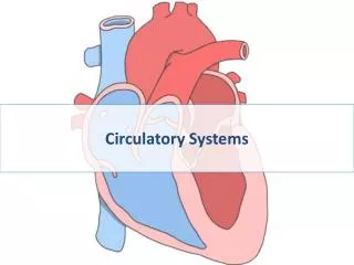
Mammaliancirculation systemic pulmonary systemic What do bluevs.redareas represent?
Mammalian heart to neck & head& arms Coronary arteries
Coronary arteries bypass surgery
Heart valves SL AV AV • 4 valves in the heart • flaps of connective tissue • prevent backflow • Atrioventricular (AV) valve • between atrium & ventricle • keeps blood from flowing back into atria when ventricles contract • “lub” • Semilunar valves • between ventricle & arteries • prevent backflow from arteries into ventricles while they are relaxing • “dub”
The heart sounds heard with a stethoscope are caused by the closing of the valves. (Even without a stethoscope, you can hear these sounds by pressing your ear tightly against the chest of a friend—a close friend.) The sound pattern is “lub–dup, lub–dup, lub–dup.” The first heart sound (“lub”) is created by the recoil of blood against the closed AV valves. The second sound (“dup”) is the recoil of blood against the semilunar valves.
Lub-dub, lub-dub • Heart sounds • closing of valves • “Lub” • recoil of blood against closed AV valves • “Dub” • recoil of blood against semilunar valves • Heart murmur • defect in valves causes hissing sound when stream of blood squirts backward through valve SL AV AV
Cardiac cycle 110 ____ 70 systolic ________ diastolic pump(peak pressure) _________________ fill(minimum pressure) • 1 complete sequence of pumping • heart contracts & pumps • heart relaxes & chambers fill • contraction phase • systole • ventricles pumps blood out • relaxation phase • diastole • atria refill with blood
The control of heart rhythm Pacemaker generates wave of signals to contract. Signals pass to heart apex. Signals are delayed at AV node. Signals spread throughoutventricles. Bundlebranches AV node SA node(pacemaker) Purkinjefibers Heartapex ECG 1 2 3 4
The cardiac cycle 2 Atrial systole; ventricular diastole Semilunarvalvesclosed 0.1 sec Semilunarvalvesopen 0.3 sec 0.4 sec AV valveopen AV valveclosed 1 Atrial and ventricular diastole 3 Ventricular systole; atrial diastole
Measurement of blood pressure Blood pressure Reading: 120/170 Pressurein cuff above120 Pressurein cuff below 120 Pressurein cuff below 70 Rubber cuffinflatedwith air 120 120 70 Sounds stop Sounds audible instethoscope Artery Arteryclosed • High Blood Pressure (hypertension) • if top number (systolic pumping) > 150 • if bottom number (diastolic filling) > 90
The composition of mammalian blood Plasma 55% Cellular elements 45% Constituent Major functions Functions Cell type Numberper L (mm3) of blood Solvent for carrying other substances Water Erythrocytes(red blood cells) Transport oxygenand help transportcarbon dioxide Icons (blood electrolytes 5–6 million Sodium Potassium CalciumMagnesium Chloride Bicarbonate Osmotic balance pH buffering, and regulation of membrane permeability Separatedbloodelements Leukocytes(white blood cells) Defense andimmunity 5,000–10,000 Plasma proteins Osmotic balance, pH buffering Clotting Defense Albumin Fibringen Immunoglobulins (antibodies) Lymphocyte Basophil Eosinophil Substances transported by blood Neutrophil Monocyte Nutrients (such as glucose, fatty acids, vitamins) Waste products of metabolism Respiratory gases (O2 and CO2) Hormones Platelets 250,000400,000 Blood clotting
Differentiation of blood cells Pluripotent stem cells(in bone marrow) Lymphoidstem cells Myeloidstem cells Basophils B cells T cells Lymphocytes Eosinophils Neutrophils Erythrocytes Platelets Monocytes
Blood clotting 2 1 3 This seal is reinforced by a clot of fibrin when vessel damage is severe. Fibrin is formed via amultistep process: Clotting factors released fromthe clumped platelets or damaged cells mix withclotting factors in the plasma, forming an activation cascade that converts a plasma proteincalled prothrombin to its active form, thrombin.Thrombin itself is an enzyme that catalyzes the final step of the clotting process, the conversion of fibrinogen to fibrin. The threads of fibrin become interwoven into a patch (see colorized SEM). The platelets form a plug that provides emergency protection against blood loss. The clotting process begins when the endothelium of a vessel is damaged, exposing connective tissue in the vessel wall to blood. Platelets adhere to collagen fibers in the connective tissue and release a substance that makes nearby platelets sticky. Collagen fibers Red blood cell Fibrin clot Plateletplug Platelet releases chemicalsthat make nearby platelets sticky Clotting factors from: Platelets Damaged cells Plasma (factors include calcium, vitamin K) Thrombin Prothrombin Fibrinogen Fibrin 5 µm
Atherosclerosis Smooth muscle Connective tissue Plaque Endothelium (a) Normal artery (b) Partly clogged artery 50 µm 250 µm
Make sure you can do the following: • Label all parts of the mammalian heart and diagram blood flow through it. • Explain the causes of circulatory system disruptions and how disruptions of the circulatory system can lead to disruptions of homeostasis.