Download
1 / 19
190 likes | 311 Views
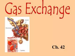
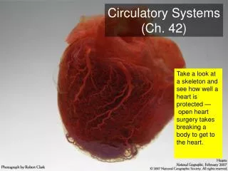
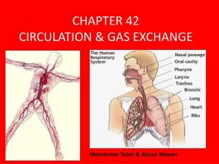
Ch 42 Circulation. AP Biology. Heart Pumps Blood. 1. Pumping of heart keeps blood moving in arteries. 2. Skeletal muscle contraction is responsible for blood movement in veins. This is much like squeezing a tube of tooth paste to get the toothpaste out of the tube.

E N D
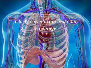
Ch 42 Circulation AP Biology
Heart Pumps Blood 1. Pumping of heart keeps blood moving in arteries. 2. Skeletal muscle contraction is responsible for blood movement in veins. This is much like squeezing a tube of tooth paste to get the toothpaste out of the tube. 3. Heart is a cone-shaped, muscular organ about size of a fist. 4. It is located between lungs directly behind the sternum and is tilted so that the apex is directed to left.
Heart Pumps Blood 5. Myocardium is major portion of the heart consisting mostly of cardiac muscle; muscle fibers are branched and tightly joined together. 6. Heart lies within a pericardium sac that contains pericardial fluid which provides cushioning. 7. Endocardium lines inner surface of the heart; it consists of connective tissue and endothelial tissue. 8. Internal wall called the septum separates heart into right and left halves.
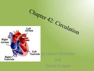
Heart Pumps Blood 9. Heart has two upper, thin-walled atria and two lower, thick-walled ventricles. a. Atria receive blood from venous portion of cardiovascular system. b. Atria are much smaller and weaker than muscular ventricles but hold the same volume of blood. c. Ventricles pump blood into arterial portion of cardiovascular system.
The Heart Structure Atrioventricular valves between atria and ventricles prevent back flow from ventricle to atrium. Right atrioventricular (tricuspid) valve on right side of heart consists of three cusps or flaps. Left atrioventricular (bicuspid or mitral) valve on left side consists of two cusps or flaps. Semilunar valves resembling half-moons are located between a ventricle and an artery that prevents back flow from artery to ventricle. 1. The pulmonary semilunar valve lies between the right ventricle and the pulmonary trunk. 2. The aortic semilunar valve lies between the left ventricle and the aorta.
Path of Blood Through the Heart Route of blood through heart is as follows. a. Deoxygenated blood enters right atrium from both superior vena cava and inferior vena cava. b. Right atrium sends blood through right atrioventricular (tricuspid) valve to right ventricle. c. Right ventricle sends blood through pulmonary semilunar valve into pulmonary trunk and arteries to lungs. d. Oxygenated blood returns from lungs through pulmonary veins and is delivered to left atrium.
Path of Blood Through the Heart e. Left atrium sends blood through left atrioventricular (bicuspid or mitral) valve to left ventricle. f. Left ventricle sends blood through aortic semilunar valve into aorta and to body.
The Heartbeat 1. Heart contracts (beats) about 70 times a minute; each heartbeat lasts about 0.85 seconds. 2. Heartbeat or cardiac cycle consists of phases: systole refers to contraction of heart chambers and diastole is relaxation of heart chambers. 3. Atria contract first while ventricles relax (0.15 sec.), then ventricles contract while atria relax (0.30 sec.), then all chambers rest (0.40 sec.). 4. Heart is in diastole about 50% of the time.
The Heartbeat 5. Short systole of the atria is needed only to send blood into ventricles. 6. When the term "systole" is used alone, it refers to left ventricle systole. 7. When the heart beats, familiar lub-dub sound is heard as valves of heart close. a. Lub is caused by vibrations of the heart when atrioventricular valves close. b. Dub is heard when vibrations occur due to closing of semilunar valves.
BLOOD PRESSURE 1. Systolic pressure results from blood being forced into arteries during ventricular systole. 2. Diastolic pressure is pressure in arteries during ventricular diastole. 3. Human blood pressure is measured as force pushing against wall of brachial artery of upper arm. a. Blood pressure is measured by a sphygmomanometer which has a pressure cuff. b. Clinical blood pressure measures pressures produced by contraction and relaxation of right ventricle. c. It is stated in millimeters of mercury (e.g., 120/80 mm Hg for systolic/diastolic). 4. As blood flows from aorta into arteries and arterioles, blood pressure falls.
Cardiovascular Disease 1. Cardiovascular disease (CVD) is the leading cause of untimely deaths in the United States. 2. Risk of CVD can be reduced by following guidelines for a heart-healthy life-style.
Hypertension 1. An estimated 20% of Americans suffer from hypertension or high blood pressure. 2. Women have this condition if their blood pressure is significantly higher than 160/95; men under age 45 if over 130/90, and beyond age 45 if above 140/95. 3. Diastolic pressure is emphasized when medical treatment is considered. 4. Hypertension may not be detected until a stroke or heart attack occurs. 5. Genetics is known to play a role in hyper-tension.
Atherosclerosis 1. Hypertension is seen in individuals with atherosclerosis, formerly called arteriosclerosis. 2. Soft plague masses of fatty materials accumulate beneath inner linings of arteries. 3. As plaque accumulates, it protrudes into a vessel, interfering with blood flow. 4. Atherosclerosis develops in early adulthood but symptoms may not appear until age 50 or older. 5. Plaque can cause a blood clot to form on irregular arterial walls. 6. A clot may remain stationary or dislodge and move through the blood. 7. In some families, atherosclerosis is inherited.
Stroke 1. Stroke, heart attack, and aneurysm are associated with hypertension and atherosclerosis. 2. Strokes can result in paralysis or death; a small arteriole bursts or is blocked by an embolus. a. Stroke is also called a cardiovascular accident (CVA). b. Paralysis or death depends on extent a portion of the brain lacks O2. c. Warning symptoms include: numbness in hands or face, difficulty speaking, blindness in one eye.
Heart Attack (MI) 1. A myocardial infarction (MI) is also called heart attack. a. It occurs when a portion of heart muscle dies due to a lack of O2. b. A partially blocked coronary artery causes angina pectoris causing chest pains or radiating pain in left arm. c. Nitroglycerin and related drugs dilate blood vessels and relieve pain. d. One cause of heart attacks is blockage of coronary arteries.
REVIEW • The heart receives oxygen-deficient blood (see the white arrows) from the body into the right upper atrium. • When the heart contracts, the right lower ventricle will pump the blood into the lungs, where the carbon dioxide is exchanged for oxygen. • After the exchange, the blood containing fresh oxygen flows into the left upper atrium. • Oxygen-rich blood (see the black arrows) flows from the left upper atrium into the left lower ventricle. • When the heart contracts, the left lower ventricle will force the blood out to the body through a network of arteries.
Common Misconception • Human blood is red, ranging from bright red when oxygenated to dark red when not. It owes its color to hemoglobin, a metalloprotein compound containing iron in the form of heme, to which oxygen binds. There exists a popular misconception that deoxygenated blood is blue and that blood only becomes red when it comes into contact with oxygen. Blood is never blue, but veins appear blue because light is diffused by skin. Moreover, the blood inside is dark red and exhibits poor light reflection. From a physiological perspective, veins and arteries appear similar when skin is removed and are seen directly.