Download
1 / 23
290 likes | 801 Views
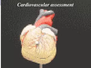
Cardiovascular Assessment. Heart and Circulation. Location and Shape Precordium Base Apex Great Vessels of the Heart Superior and Inferior Vena Cava Pulmonary Artery Pulmonary Veins Aorta. Heart Structure. Layers Pericardium Myocardium (subendocardium) Endocardium Valves Semilunar

E N D
Heart and Circulation • Location and Shape • Precordium • Base • Apex • Great Vessels of the Heart • Superior and Inferior Vena Cava • Pulmonary Artery • Pulmonary Veins • Aorta
Heart Structure • Layers • Pericardium • Myocardium (subendocardium) • Endocardium • Valves • Semilunar • Aortic, Pulmonic • Atriventricular • Bicuspid (Mitral), Tricuspid
Conductance System • SA node • AV node • Bundle of HIS • Purkinje fibers • Myocardium
Cardiac Cycle • Diastole • Early filling (passive) • Presystole (Atrial kick) – 25% of stroke volume • Systole • AV valves shut (S1) • Pressure builds, shoots through SL valves • SL valves shut (S2)
Heart Sounds • Normal • S1 • S2 • Splitting • Extra • S3 • S4 • Murmur
Murmur • Causes • Increased blood velocity • Decreased blood viscosity • Structural defects, especially valve and septal • Grading: 1 – 6
Pumping action • Preload – volume of blood in heart before systole; also end-diastolic volume (EDV) • Afterload – diastolic blood pressure; pressure that the heart must overcome to pump blood • Stroke Volume (SV) – volume of blood pumped in one heartbeat • Ejection Fraction – SV/EDV
Developmental Concerns • Infants – foramen ovale, ductus arteriosus • Pregnant – Increase in blood volume • Aging adult • Systolic pressure increases • Myocardial thickening • Decreased exercise augmentation
History • Chest Pain • Acute • Angina pectoris • Stable • Unstable • Dsypnea • Paroxsymal • Orthopnea
History • Cough • Hemoptysis • Frothy pink sputum • Fatigue • Cyanosis, pallor • Edema • Nocturia
History • Past cardiac history • MI, HTN, cholesterol, murmur, congenital disease, rheumatic fever, valve problem, DM • Family history • HTN, cholesterol, CAD, DM, CVA • Personal habits • Nutrition, Smoking, ETOH, Exercise, Drugs
Physical Exam • Inspect chest • Palpate Apical pulse • Palpate precordium for thrills • Percussion for heart size • Auscultation • Rate • Rhythm • Murmurs, Rubs
Lub Dub • S1 heard first • S2 heard second • Exception - Tachycardia • Splitting • Abnormal • S3 – fluid overload: HF • S4 – ventricular resistance (hardened ventricle)
Heart Failure Findings • Daily weight • BP • HR • Orthopnea, PND • S3 • Breath Sounds • DOE Stages 1 – 4
Inspect: JVD Auscultate Temporal Carotid Aorta Renal Iliac Femoral Palpate Carotid Brachial Radial Ulnar Femoral Popliteal Posterior Tibial Dorsalis Pedis Peripheral Vascular System
Other • Veins • Know difference between deep and superficial veins in arms and legs. Know what perforators do. • Homan’s sign • Skin • Color, warmth • Edema
Vocabulary • Doppler ultrasound • Peripheral lymph nodes to palpate • Epitrochlear, axillary, femoral • Raynaud • Lymphedema • Ischemic ulcer • Venous stasis ulcer • Varicose veins • DVT • PAD, PVD