Download
1 / 34
340 likes | 483 Views
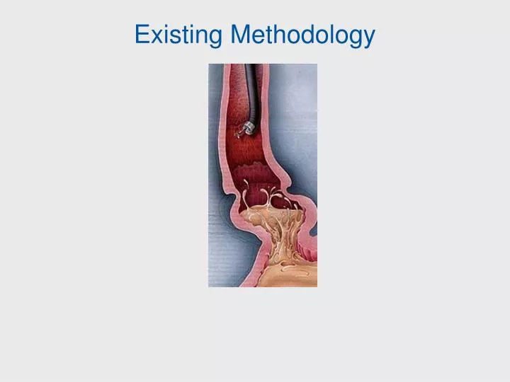
Existing Methodology. Question: Will we be able to do this?. Question: What is “normal” tissue?. Surrogate Anatomic Sites for Evaluating Cancer Risk. Vadim Backman, Ph.D. Biomedical Engineering Department Northwestern University. Colorectal Cancer (CRC).

E N D
Surrogate Anatomic Sites for Evaluating Cancer Risk Vadim Backman, Ph.D. Biomedical Engineering Department Northwestern University
Colorectal Cancer (CRC) • #2 cause of cancer deaths, ~155K cases annually and ~57K deaths – stagnant for 20 years • 90%+ survival rate if caught early but today the majority (61%) are later stage • Colonoscopy detects and removes a precursor to colon cancer, adenomatous polyp, thus decreasing future occurrence of CRC by 75-90%
Why Colon Cancer Screening Program Does Not Work? • Problem: • Current guidelines: everybody over age 50 is recommended to undergo colonoscopy at least once every 10 years. • There are >90 million Americans over age 50. • 70-80% of colonoscopies are negative and unnecessary • Colonoscopic screening of this entire eligible population is impossible due to • - expense (annual cost would be ~$50 billion) • - insufficient number of endoscopists • - patient reluctance (hate prep!) • - complication rate. • 85% of the population receives no colonoscopic CRC screening. • Solution: Develop a minimally invasive technology to identify patients who are at risk for CRC and would benefit from colonoscopy. Precedent: Cervical cancer screening with Pap smear in the last 50 years reduced mortality from #1 cancer in women to #13!
Bowel Bowel Technique Technique Expensive? Expensive? preparation preparation Sensitivity* required? required? 100%** Colonoscopy Colonoscopy Yes, >$1,000 Yes, >$1,000 Yes Yes FOBT FOBT No No No No 10% Fecal DNA Fecal DNA Yes, $700 No No 18% 18% Virtual Virtual 55-80% Yes, ~$1,000 Yes Yes colonoscopy colonoscopy Imaging Imaging Not yet approved Not yet approved Yes, >$2,000 Yes, >$2,000 Yes Yes capsule capsule for colon imaging for colon imaging Factors for A Successful Screening Test: Inexpensive, No Prep, & Sensitive • Requirements for a population screening test: • No bowel preparation • Performed by a PCP • Sensitive • Inexpensive *Sensitivity relative to colonoscopy for precancerous advanced adenomas (polyps >1cm). Precancerous polyps, not cancer, is the most clinically relevant for the screening population. **Gold standard with assumed sensitivity of 100%. Colonoscopy is estimated to miss up to 10% of advanced lesions (>1cm) and up to 35% of smaller lesions
Exploiting the Field Effect Field Effect: the genetic/environmental milieu that results in a neoplastic lesion in one area of the colon exists throughout the organ. • Conventional wisdom: tissue in a tumor is abnormal, tissue surrounding tissue is normal. • This is only an approximation • New methodology: detection of carcinogenesis by analysis of normal appearing cells in an accessible part of an organ.
Commonly Used Biomarkers of Field Effect in CRC • Focal Neoplastic Lesions to predict Proximal Neoplasia • Adenoma on flexible sigmoidoscopy (Arch Intern Med. 2004 1881-7). • Rectal aberrant crypt foci (Takayama et al N Engl J Med. 1998; 339:1277.) • Diffuse Alterations in the Histologically Normal Mucosa • Decreased apoptosis (Bernstein et al. Cancer Res. 1999 59:2353-7) • Increased proliferation (Ponz de Leon, et al Cancer Res. 1988 ;48:4121).
Novel Biomarkers of Field Effect in CRC Microarray evidence Proteomic evidence TUMOR Polley et al. Cancer Res 2006 Chen et al. Cancer Res 2004
Optically-detectable Markers of the Field Effect • Our approach: sensing changes in tissue that CANNOT be detected by histopathology, spatially outside the extent of a neoplastic lesion. • Tissue physiology: Increased mucosal microvascular blood supply • Tissue morphology: Alterations in tissue fractal microarchitecture • Intracellular morphology: Alterations in intracellular nanoscale architecture
Lessons from Animal Studies • AOM-treated rat and MIN-mouse models • Early increase in microvascular blood supply (EIBS) and alterations in tissue micro and nano-architecture develop prior to ACFs and microadenomas. • These alterations can be detected at a distance from the neoplastic focus. • Diffusely present in ~90% of tissue sites. • Depth-resolution is crucial: changes are only in the mucosa (top 100 m). • EIBS is caused in part by iNOS upregulation. Gastroenterology, 126, 1071-1081 (2004) Clinical Cancer Research, 19, 961-968 (2006) FEBS, 581, 3857-3862 (2007) Gut, 54, 654-660 (2005)
Optically-detectable Markers of the Field Effect • Tissue physiology: Increased microvascular blood supply • Tissue morphology: Alterations in tissue microarchitecture • Intracellular morphology: Alterations in intracellular nanoscale architecture
EIBS: In Vivo Clinical Validation • Technology: polarization-gated spectroscopy sensitive to mucosal microcirculation • Design: in vivo, during colonoscopy • Patient characteristics: • 220 average risk screening patients • 51 with adenomas: 30 non-advanced adenomas, 9 multiple non-advanced adenomas,12 advanced • 169 patients with no neoplasia including 26 with hyperplastic polyps colonoscope EIBS fiber-optic probe
Rectal EIBS is Indicative of Presence of Adenomas Throughout the Colon * * Patients with no dysplasia vs. patients with advanced adenomas Area under ROC curve =90% Rectal EIBS EIBS reading: rectum only Adenoma location: throughout the colon Sensitivity = 100% Specificity = 75%
Optically-detectable Markers of the Field Effect • Tissue physiology: Increased microvascular blood supply • Tissue morphology: Alterations in tissue (fractal) microarchitecture • Intracellular morphology: Alterations in intracellular nanoscale architecture
Alterations in Mucosal Microarchitecture: Clinical Study Results • Technology: low-coherence enhanced backscattering (LEBS) spectroscopy • Design: rectal biopsy • Patient characteristics: • 233 patients undergoing screening colonoscopy • Mean age 56.8 ±10.7 • 47% female • 60 with adenomas (17 advanced adenomas) • 9 with previous h/o adenomas but none on present colonoscopy • 158 with no current, prior or family history of adenomas
Potential Confounding Factors Do Not Appear to Affect LEBS Diagnosis
Optically-detectable Markers of the Field Effect • Tissue physiology: Increased microvascular blood supply • Tissue morphology: Alterations in tissue microarchitecture • Intracellular morphology: Alterations in cell nanoscale architecture
1 µm 1 µm Alterations in Epithelial Nanoarchitecture: Clinical Study Results • Technology: partial wave spectroscopic (PWS) microscopy • Design: rectal mucosal brushings • Patient characteristics: • 35 patients • 21 with no neoplasia • 14 with adenomas, 4 advanced
x 10-7 13 * 12 * p-value < 0.0001 11 Disorder strength Ld (mm) * 10 9 8 7 Adenoma Advanced Control adenoma Human Clinical Study Results no neoplasia neoplasia Disorder in nanoscale density fluctuations in endoscopically-normal rectal mucosa is increased in patients with sporadic adenomas
Colonoscopy-free Screening for Colon Cancer Using Optical Detection of the Field Effect Annual population screening by PCP’s during an annual exam without colonoscopy and preparation _ + Colonoscopy • Only patients with adenomas receive colonoscopies • Most (all) patients with adenomas are screened • Patients are more compliant • Better allocation of colonoscopic resource LEBS probe
Does LEBS Work in Other Organs? Example: Pancreatic Cancer • No existing technique is capable of accurate diagnosis of pancreatic carcinogenesis in preinvasive (PanIN) or resectable stage. • 95% mortality within a year after diagnosis. • Problem: pancreatic duct exam is not suitable for screening due to a high rate of complications including acute pancreatitis (~5-20%). • Solution: PWS analysis of duodenal periampullary cells brushed during upper endoscopy.
Prospect of Pancreatic Cancer Screening PWS (Nanoarchitecture): LEBS (Microarchitecture): 204 patients total 84 Healthy control 26 Family History 29 Cyst 45 Pancreatic Adenocarcinoma 20 Other diseases 35 patients total 26 Healthy control 9 Pancreatic Adenocarcinoma Testing set: Sensitivity = 85% Specificity = 80% Sensitivity = 90% Specificity = 81%
Example III: Lung Cancer • >80% of lung cancer patients had altered nuclear texture features • Us-Krasovec et al., Anal Quant Cytol Histol. 2005 Oct;27(5):254-62 • Automated Quantitative cytology of buccal nuclei correlated with lung cancer • 66% sensitivity and 70% specificity for lung cancers • 61% sensitivity for stage 1 • Turic et al, Chest 2005 (abstract) • Increased incidence of head and neck cancer in patients with lung cancer • Johnson et al., B. J. Natl. Cancer Inst., 1998 • Genetic changes in the histologically normal large-airway epithelial cells obtained at bronchoscopy. • Guo, M. et al. Clin. Cancer Res. 2004. • 80% sensitivity and 84% specificity • Spira et al., Nat Med 2006.
Lung Cancer Screening by PWS Analysis of Buccal Cells • Normal - Nonsmokers - 5 • Cancer - Nonsmokers - 3 • COPD (smokers) - 31 • COPD – Family Hx - 7 • Lung cancer (smokers) - 53 • Other cancers - 4 • Smokers (no COPD or cancer) - 5 Number of Patients: 108
PWS Images are Different for Non-cancer and Cancer Patients Bright field Non-cancer (COPD) patients PWS Disorder strength (Ld), um Bright field Lung cancer patients PWS
P<0.001 Control (COPD) Lung cancer Disorder in Nanoarchitecture is Increased in Buccal Cells in Lung Cancer Patients Area under ROC curve = 84% Sensitivity = 90% Specificity = 77%
Potential Confounding Effects • Can the differences in cell nanoarchitecture be simply due to difference in age among COPD and lung cancer patients? • Can the differences be due to different smoking history?
Conclusions • What we call “histologically normal tissue” is not entirely normal in patients with neoplasia • Not only neoplastic lesions but also tissue outside neoplastic lesions is abnormal • Biophotonics can detect field effects associated with carcinogenesis in the colon, pancreatic and lung • Optically-detectable markers of the field effect include increased mucosal blood supply, micro and nano-architectural changes in the mucosa • Potential for colon, pancreatic and lung cancer screening/risk-stratification through biophotonics detection of the field effect
Northwestern University Vladimir Turzhitsky Andrew Gomez Hariharan Subramanian Sarah Ruderman Jeremy Rogers, Ph.D. Young Kim, Ph.D. Yang Liu, Ph.D. Prabhakar Pradhad, Ph.D. Xu Li, Ph.D. Alexei Kromine Acknowledgements Funding National Institutes of Health R01CA128641, R01 CA109861, R01 EB003682, R01 CA112315, R01 CA118794, R33CA122017, R21 EB006742, U01 CA11125 National Science Foundation CBET- 0733868, CBET-0238903 V Foundation Coulter Foundation AACR Evanston Hospital Hemant Roy, M.D. Ramesh Wali, Ph.D. Randall Brand, M.D. M. Goldberg, M.D.