Download
1 / 58
610 likes | 762 Views
Cell Signaling and Communication. Cell Signaling and Communication. Signals Receptors Signal Transduction Signal Effects: Changes in Cell Function Direct Intercellular Communication. Signals.

E N D
Cell Signaling and Communication • Signals • Receptors • Signal Transduction • Signal Effects: Changes in Cell Function • Direct Intercellular Communication
Signals • Both prokaryotic and eukaryotic cells must process information from their environment and respond appropriately. • Signals may be chemical molecules or physical stimuli such as light. • Cells must be set up to interpret signals—not all cells can interpret all signals. • To interpret a signal, a cell must have the appropriate receptor protein.
Signals • Organisms receive many signals from the environment such as light, odors, tastes, temperature, touch, and sound. • Multicellular organisms’ internal cells are exposed to extracellular fluids and other cells, from which they receive information. • A few of the many types of signals in animal cells are hormones, neurotransmitters, chemical messages from the immune system, CO2, and H+.
Signals • In large animals, signals reach targets via diffusion as autocrine or paracrine signals when the target is close. • Autocrine signals are signals generated by the same cells upon which they act. • Paracrine signals diffuse to and affect nearby cells. • When the target is distant, signals travel by circulation in the blood.
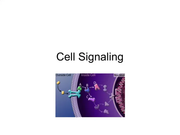
Signals • The entire signaling process, from signal detection to final response, is called a signal transduction pathway. • A signal transduction pathway involves a signal, a receptor, transduction, and effects. • It is usually described as a series of events but many of the events are actually happening at the same time.
Signals • An example using E. coli: • The signal is rising solute concentration outside the cell. • The receptor protein is EnvZ, a transmembrane protein. Rising solute concentration changes the protein’s conformation. • EnvZ becomes a kinase, and phosphorylates itself. • A responder is the second component in the pathway. EnvZ now binds to OmpR, which takes the phospate group. OmpR changes shape.
Signals • The signal on the outside of the cell has been transduced to a protein inside the cell, the phosphorylated OmpR. • Phosphorylated OmpR is a transcription factor. It binds to the promoter for the ompC gene. • The protein OmpC is inserted into the outer membrane where it blocks pores and prevents solutes from entering.
Signals • A review of the steps in this signal transduction pathway: • A receptor binds with the signal molecule and changes shape. • Conformational change results in kinase activity. • Phosphorylation alters the functioning of a protein. • The signal is amplified. • Transcription factors are activated. • Altered synthesis of specific proteins occurs. • Protein action alters cell activity.
Receptors • A cell responds to only a few of the many signals it receives. • The type of receptors each cell makes is genetically determined. • Receptors have specific binding sites for their signals.
Receptors • A ligand is the signaling molecule that binds the receptor. • Binding of the ligand causes the receptor to change shape. • The ligand has no further involvement in the pathway. • Receptors bind ligands according to the law of mass action, and thus the binding is reversible.
Receptors • Inhibitors can bind to the ligand binding sites on receptor molecules. • Natural and artificial inhibitors are important in medicine.
Receptors • There are two classes of signaling molecules: • Ligands with cytoplasmic receptors: small and/or nonpolar molecules that can cross the plasma membrane, such as steroids. • Ligands with plasma membrane receptors: large and/or polar molecules that can not cross, such as insulin. Receptors are usually transmembrane proteins.
Receptors • Three well-studied types of transmembrane receptors in complex eukaryotes: • Ion channel receptors • Protein kinases • G protein-linked receptors
Receptors • Some ion channel proteins, acting as “gates,” are signal receptors. • Channel proteins can open to let certain ions in or out, or close to restrict them. • The signal to open or close the channel can be chemical, light, sound, pressure, or voltage. • An example of a gated ion channel is the acetylcholine receptor.
Receptors • Some eukaryotic receptor proteins become kinases when activated. • A phosphate is transferred from ATP to a protein, the target protein, changing its shape or activity. • Sometimes the protein kinase phosphorylates itself. This is called autophosphorylation. • Insulin receptors are examples of protein kinase receptors.
Receptors • The seven-spanning G protein-linked receptors are proteins with seven regions that pass through the lipid bilayer. • A ligand binds to the extracellular side and changes the shape of the protein on the cytoplasmic side. This exposes a binding site for the G protein. • G protein also has a binding site for GTP. The GTP-bound subunit separates and moves along the membrane until it finds an effector protein. • The effector protein may catalyze many reactions, amplifying the signal.
Receptors • G proteins can either activate or inhibit effectors. Epinephrine illustrates both possibilities. • In the heart, epinephrine causes the G protein to activate an enzyme that produces cAMP, which has a wide range of effects on the cell. • In smooth muscle cells around blood vessels, epinephrine causes the G protein to inhibit the production of cAMP, muscles relax, and the blood vessels open wide for maximum blood flow.
Receptors • Cytoplasmic receptors which are located inside the cell bind with ligands that can cross the plasma membrane. • The receptor changes shape and can then enter the nucleus where it acts as a transcription factor. • Steroid hormones are an example of such signal molecules.
Signal Transduction • Transducers convert signals from one form to another. • Direct transduction results from the action of the receptor itself on effector proteins. Direct transduction occurs at the plasma membrane. • Indirect transduction uses a second messenger to mediate the interaction between receptor binding and cellular reaction. • In both direct and indirect transduction the signal initiates a series of events that eventually lead to a final response.
Signal Transduction • A protein kinase cascade is direct signal transduction that catalyzes the phosphorylation of target proteins. • Details of a certain protein kinase cascade were discovered from the investigation of Ras protein inhibition as treatment for bladder cancer. • Ras is part of a protein kinase cascade that influences cell division. The pathway is called a cascade because each kinase phosphorylates the next.
Signal Transduction • There are at least three advantages to having many kinase steps in signal transduction: • Each activated protein kinase can phosphorylate many target proteins, so amplification of the signal occurs at each step. • A signal at the cell membrane is transferred to the nucleus. • Having many steps affecting different target proteins allows for a variety of responses by different cells to the same signal.
Signal Transduction • Indirect transduction is more common than direct transduction. • Scientists investigating the effects of epinephrine on the liver enzyme phosphorylase discovered cyclic AMP (cAMP) as a second messenger. • The second messenger carries the signal from the membrane receptor to the cytoplasm. • Second messengers affect many cell processes, amplifying the signal.
Signal Transduction • The cAMP molecule is a small cyclic nucleotide generated from ATP. • The enzyme adenylyl cyclase produces cAMP using ATP as a substrate. Adenylyl cyclase is activated by an activated G protein subunit. • Like other second messengers, cAMP is not an enzyme. Second messengers act as cofactors or allosteric regulators of target proteins. • cAMP has two major kinds of targets: ion channels and protein kinases.
Signal Transduction • Phospholipids can be hydrolyzed into components that act as second messengers. • Phosphatidyl inositol-bisphosphate (PIP2) is hydrolyzed into inositol triphosphate (IP3) and diacylglycerol (DAG). • The two parts each become second messengers, with IP3 moving into the cytoplasm and DAG remaining in the membrane. • These second messengers trigger many cellular events.
Signal Transduction • Calcium ions are also second messengers. • Ca2+ concentration in the cytoplasm is usually only about 0.1 mM. • The concentration is kept low via active transport, both out of the cell and into the ER. • Unlike cAMP, Ca2+ cannot be manufactured in the cell; it must be imported. • Many different signals cause Ca2+ channels to open, including IP3.
Signal Transduction • Once a signal triggers Ca2+ channels to open, Ca2+ concentration rapidly rises to 100 times the resting concentration. • The calcium ions then affect the activities of cellular proteins, including protein kinase C. • Ca2+ also binds to Ca2+ channel proteins, triggering additional releases of Ca2+. • Calcium ions bind to a calcium-binding protein called calmodulin, which can activate certain proteins.
Signal Transduction • The gas nitric oxide (NO) was found to be a second messenger by scientists studying the effects of acetylcholine, which causes the relaxation of smooth muscles of the blood vessels. • Acetylcholine stimulates the IP3 pathway to produce an influx of Ca2+, which leads to an increase in the level of another second messenger, cGMP. • This messenger stimulates a kinase cascade leading to muscle relaxation.
Signal Transduction • However, the pathway does not work in isolated artery tissue, which lacks an endothelial lining. • It was discovered that NO, produced by the endothelial cells, was also needed. • Acetylcholine causes increased Ca2+ levels in the endothelial cells, which causes the activation of NO synthase, the enzyme that makes NO. • NO diffuses rapidly from the endothelial cells to the nearby smooth muscle cells. • In the smooth muscle cells, NO activates the enzyme guanylyl cyclase, which stimulates the formation of cGMP.
Signal Transduction • Cells must regulate the activity of transducers. • NO is unstable and breaks down quickly, so NO is regulated by how much of it is made. • Ca2+ concentrations are restored by mechanisms such as membrane pumps and ion channels. • Protein kinase cascades are interrupted by protein phosphatases that remove the added phosphates, deactivating the kinases. • GTPases deactivate G proteins by converting GTP to GDP. • Both cAMP and cGMP are converted to AMP and GMP by their respective phosphodiesterases.
Signal Effects: Changes in Cell Function • Signal effects may include: • The opening of membrane channels • Changes in enzyme activity • Differences in gene transcription
Signal Effects: Changes in Cell Function • Sensory nerve cells of the sense organs are stimulated through the opening of ion channels. • Each of the thousands of nerve cells in the nose expresses just one of these receptors. • When an odorant molecule binds to its receptor, a G protein becomes activated, which leads to formation of the second messenger, cAMP. • The cAMP binds to ion channels, causing them to let in Na+. • The change in Na+ ion concentration stimulates the neuron to send a signal to the brain.
Figure 15.14 A Signal Transduction Pathway Leads to the Opening of Ion Channels (Part 1)
Figure 15.14 A Signal Transduction Pathway Leads to the Opening of Ion Channels (Part 2)
Signal Effects: Changes in Cell Function • The effects of epinephrine on liver cells results in altered enzyme activity. • The binding of epinephrine to a G protein-linked receptor results in synthesis of cAMP, which in turn initiates a series of kinase reactions. • Two enzymes are altered: • Glycogen synthase is deactivated by phosphorylation. • Glycogen phosphorylase is activated, catalyzing the release of glucose molecules from glycogen.