Download
1 / 42
450 likes | 594 Views
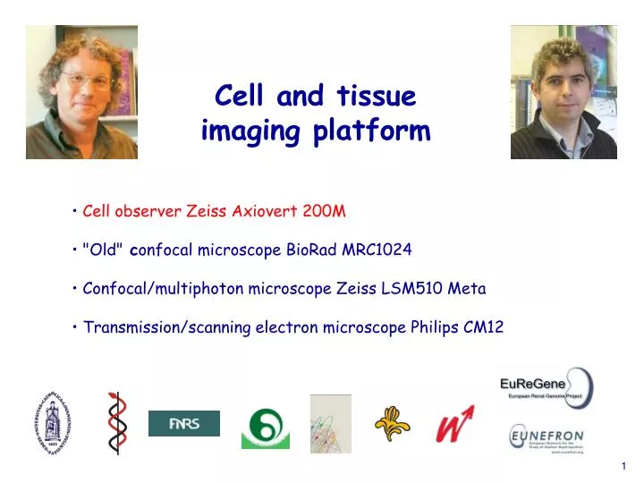
Cell and tissue imaging platform. Cell observer Zeiss Axiovert 200M "Old" c onfocal microscope BioRad MRC1024 Confocal/multiphoton microscope Zeiss LSM510 Meta Transmission/scanning electron microscope Philips CM12. Cell Observer Zeiss Axiovert 200M. Applications : General structure

E N D
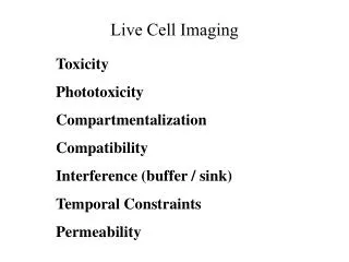
Cell and tissue imaging platform • Cell observer Zeiss Axiovert 200M • "Old" confocal microscope BioRad MRC1024 • Confocal/multiphoton microscope Zeiss LSM510 Meta • Transmission/scanning electron microscope Philips CM12
Cell Observer Zeiss Axiovert 200M • Applications : • General structure • Conventional fluorescence microscopy • Time-lapse of slow movement • Rapid movement • Image analysis (~ Metamorph) • Accessibility : free of charge (expected contribution to maintenance • costs for significant users) • Modulability
Conventional microscopy : stained sections in situ hybridization for CXCR4 (mouse embryo e12) salivary gland C. Pierreux and A.C. Hick (CELL)
Conventional microscopy : living specimens differentiation of epithelial islands Bright Field SEM W. Rezende-Lima and P. Van Der Smissen (CELL)
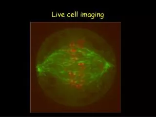
Slow movement: time-lapse regulation by Src of actin-dependent motility Src/ts inactive (40°C) Src/ts active (34°C) Platek et al, 20O4, J. Cell Sci. 117 : 4849-61
Rapid ciliary movement courtesy of Drs F. Tissir and A. Goffinet (DENE)
Image analysis platform (AxioVision and ImageJ : ~ Metamorph) • Applications : • Morphometry • - Size distribution • - Surface of complex domains • Dynamics • - Track analysis • Multiple other applications
class I class II class III 5 µm recovery after photobleaching(%) time (sec) time (sec) time (sec) Morphometry of complex domains micrometric domains of plasma membrane lipids Tyteca et al, in preparation
Cell and tissue imaging platform • Cell observer Zeiss Axiovert 200M • "Old" confocal microscope BioRad MRC1024 • Confocal/multiphoton microscope Zeiss LSM510 Meta • Transmission/scanning electron microscope Philips CM12
Confocal microscope BioRad MRC1024 Characteristics : ExcitationEmissionTypical fluorochromes 488 nm (blue) 522/35 nm (green) FITC, Alexa-488 568 nm (yellow) 605/32 nm (red) TMR, Alexa-568 647 nm (red) 680/32 nm (far red pseudocolor blue) To-Pro, Cy5 Attention ! “out of service” new user friendly equipment should be requested by a consortium in 2009
Confocal microscope BioRad MRC1024 • Applications : • Confocal microscopy: triple labelling • Time-lapse • FRAP • Thermostated stage (4°C -30°C) • Accessibility : • Free training (Patrick Van Der Smissen) general introduction to small groups • back-up for two individual sessions referenced users with private login • First come / first served • 20 EUR /h in 2008 • Methods update : testing of new reagents • Supply of unusual secondary reagents free of charge
control AICAR, 1 mM, 20 h 10 µm Triple labelling by confocal microscopy AMPK controls actin organization MDCK- I :actin(stress fibers),paxillin(focal adhesion),Topro-Blue(nuclei) Horman et al, in preparation
0 sec 9 sec 18 sec 27 sec 36 sec 45 sec 53 sec 62 sec 71 sec 80 sec 89 sec 98 sec 2 µm (fluo t – fluo bleach) (fluo initial – fluo bleach) Recovery at time t = recovery after photobleaching(%) time (sec) Lateral mobility at the plasma membrane : multiple FRAP after TMA-DPH labeling of CHO cells CTL zone ; Bleached zone A ; Bleached zone B • T1/2 recovery • mobile fraction
Cell and tissue imaging platform • Cell observer Zeiss Axiovert 200M • "Old" confocal microscope BioRad MRC1024 • Confocal/multiphoton microscope Zeiss LSM510 Meta • Transmission/scanning electron microscope Philips CM12
Confocal/multiphoton Zeiss LSM510 • Characteristics : • Increased sensitivity of PMTs (20-40 x > MRC1024) • Depth penetration (up to 400 µm) • Extended observation ( > 4 h)
Principle of multiphoton microscopy excitation by one photon at high energy is replaced by a rapid succession (10-13 sec) of 2 (or 3) photons of 1/2 (or (1/3) energy
1-Photon 2-Photons focal point 1-Photon In multiphoton microscopy, restriction of excitation to the focal point prevents photodamage above or below 2-Photons
Broad excitation and emission possibilities Excitation : • visible range : • laser Ar (458/477/488/514 nm, 30 mW) • laser DPSS (561 nm, 10 mW) • laser HeNe (633 nm, 5 mW) • infra-red range : pulse-laser (continuous) • Coherent Chameleon Ultra Emission : • nearly continuous 400 -1000 nm spectrum ; 10 nm band
Confocal/multiphoton Zeiss LSM510 + thermostated chamber (25-40°C) with CO2 • Applications : • quadruple labelling (sequential acquisition) • spectral resolution of 4 GFP variants • in-depth analysis of thick tissues and in vivo organs • time-lapse • FRAP • FRET • Accessibility : • free training on individual basis (Patrick Van Der Smissen) referenced users with private login • protected data back-up (NAS) • first come / first served • 30 EUR /h in 2008
Spectral resolution of CFP, CGFP, GFP and YFP live cell imaging of ER, nuclei, plasma membranes and mitochondria CFP CGFP GFP YFP single labeled controls CFP CGFP YFP GFP
50 µm Three-dimensional cell migration brain slices; neurons expressing Thy1-YFP Stack 450 µm x 450 µm x 150 µm
Fluid-phase endocytosis in the kidney Alexa568-dextran 10 sec
Alexa568-dextran 20 sec
Alexa568-dextran 30 sec
Alexa568-dextran 3 min
Alexa568-dextran 20 min
Receptor-mediated endocytosis and proteolysis FITC-OVA + TxRed-OVA 17 min
FITC-OVA + TxRed-OVA 23 min
FITC-OVA + TxRed-OVA 30 min
FITC-OVA + TxRed-OVA 43 min
FITC-OVA + TxRed-OVA 130 min
Acidification in the kidney by ratio-imaging injected BCECF- dextran distal urine,pH < 5 lysosomes,pH ~ 5.4 plasma, pH 7.4
2 µm Test of association : 1. co-localization ( ~ 500 nm) CD8 TC-R merge anergic CTL competent CTL P. Van Der Smissen (CELL) in collaboration with N. Demotte and P. Van der Bruggen (LICR)
Test of association : 2. co-patching (~50 nm) merge anergic CTL CD8 TC-R competent CTL P. Van Der Smissen (CELL) in collaboration with N. Demotte and P. Van der Bruggen (LICR)
Test of association : 3. FRET (~5 nm) principle ~ 5 nm excitation TC-R (Alexa 568) CD8 (Alexa 488) emission before measure after bleaching P. Van Der Smissen (CELL) in collaboration with N. Demotte and P. Van der Bruggen (LICR)
Cell and tissue imaging platform • Cell observer Zeiss Axiovert 200M • "Old" confocal microscope BioRad MRC1024 • Confocal/multiphoton microscope Zeiss LSM510 Meta • Transmission/scanning electron microscope Philips CM12
Transmission/scanning electron microscope Philips CM12 Accessibility : Collaborations
Transmission electron microscopy receptor-mediated endocytosis in kidney PTC + HRP cytochemistry B. K. Kishore et al (1996), Lab.Invest. 74 : 1013-1023
Scanning electron microscopy : thermoactivation of v-Src tsLA31 induces circular apical ruffling 2 µm Mettlen et al (2006), Traffic 7 : 589-603
Immunogoldsurface labelling ASGP receptors on hepatocytes no ligand : random + ligand : clustered Van Der Smissen et al (1992), Eur. J. Cell Biol. 60 : 122-130
Forthcoming equipments and applications : • Stereodissection microscope with fluorescence (GFP transgenic mice) • FLIM (fluorescence life time imaging) • 3D-deconvolution All suggestions for collaboration are welcome !!!