Download
1 / 29
360 likes | 885 Views
T Cell Activation. Costimulation, Inhibition, and the Immunologic Synapse. Immunology’s “Dirty Little Secret”. Early experiments demonstrated that antigen-derived (i.e. TCR-mediated) signals alone are insufficient to initiate an immune response

E N D
T Cell Activation Costimulation, Inhibition, and the Immunologic Synapse
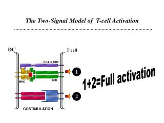
Immunology’s “Dirty Little Secret” Early experiments demonstrated that antigen-derived (i.e. TCR-mediated) signals alone are insufficient to initiate an immune response A second substance - an “adjuvant” - is required to prevent the induction of tolerance In vitro, triggering the TCR alone also leads to a tolerant state (known as “anergy”) “Two-Signal Hypothesis”: Activation of a naïve T cell requires signals from both the TCR (antigen-specific) and a second, co-stimulatory receptor (antigen-independent) Adjuvants work in part by inducing antigen presenting cells to express ligands for co-stimulatory receptors
CD28: The First (and still the champ!) Co-stimulatory Molecule Yet another member of the immunoglobulin superfamily (single Ig-V domain) Expressed on the surface of almost all T cells (100% of mouse T cells, ~80% of human T cells) as a disulfide-linked dimer Binds to B7-1 (CD80) and B7-2 (CD86) expressed on antigen presenting cells Cytoplasmic tail has binding motifs for several signaling molecules (PI3K, Grb2, Itk), but no ITAM. It is still unclear what signals CD28 contributes to T cell activation Signaling by CD28 alone does not stimulate T cells, and only activates PI3K
CTLA-4: Putting on the Brakes CTLA-4 is another CD28-related receptor, and binds both B7-1 and B7-2 - avidity is at least 20x as high as CD28 CTLA-4 expression on the surface is undetectable in resting T cells, but is rapidly increased after TCR + CD28 signaling Unlike CD28, CTLA-4 is inhibitory, and blocks T cell proliferation and IL-2 production - combination of competing B7 molecules away from CD28 and bona fide inhibitory signals (possibly phosphatases, but still not well defined)
Fig. 8.12 T-cell activation through the T-cell receptor and CD28 leads to the increased expression of CTLA-4, an inhibitory receptor for B7 molecules.
Polarization of T Cells Part I: The Cytoskeleton • T cell responses are directed at the APC/target cell, not in all directions • This requires reorganization of the cell to have a “front” (toward the APC) and a “back” - induced by signals from the TCR and costimulatory molecules • Result: Reorganization of the cytoskeleton causes reorientation of cytosolic organelles toward APC - Golgi, secretory vessicles, and microtubule organizing center (MTOC). The MTOC connects actin cytoskeletal changes with the tubulin cytoskeleton
Fig. 8.29 The polarization of T cells during specific antigen recognition
Polarization of T Cells Part II: Lipid Rafts Lipid rafts - also called GEMs (glycolipid enriched microdomains) and DIGs (detergent-insoluble glycolipid-rich domains) - are plasma membrane microdomains Enriched in cholesterol, glycolipids, and sphingolipids (i.e. lipids that are not glycerol-based), making them more rigid than the surrounding membrane Some membrane proteins are segregated - selectively enriched or depleted in rafts. Many key signaling molecules (esp. src-family kinases) are constitutively localized to lipid rafts. Dynamic structures - small rafts can condense to form larger rafts During T cell activation, TCR, CD28, and many adhesion molecules are recruited into lipid rafts, where they can interact with signaling molecules
The Immunologic Synapse - Putting it Together The combination of cytoskeletal rearrangement and lipid raft redistribution leads to the formation of a Supramolecular Activation Complex (SMAC) - a highly ordered arrangement of receptors, adhesion molecules, and signaling molecules
Effector T Cell Functions: CTL Th1/Th2 Regulatory T Cells Peripheral Tolerance Abbas Chapters 10, 13
T Cell Subsets • CD8/CTL • CD4/TH cells 1) TH1 - inflammatory response 2) TH2 - anti-inflammatory, B cell response 3) Treg - inhibit immune responses
The main function of CD8+ cells is to differentiate into CTL, which kill pathogen-infected cells
Mechanisms of Cytotoxicity • CTL (but not naïve CD8+ T cells) express lytic granules: perforin, granzymes, granulysin • Perforin lysis of target cell (inefficient) • Granzyme activation of cytosolic apoptosis machinery • Fas/FasL induction of apoptosis
CD4+ Subsets: TH1 vs. TH2 • Highly investigated in mouse model of leishmaniasis: C57BL/6 mouse cures, but Balb/c mouse dies of uncontrolled infection • Strains differ in cytokine responses • Resistant mice produce interferon-g (IFN-g), tumor necrosis factor-a (TNF-a), and lymphotoxin, which activate inflammatory response and cell-mediated immunity: TH1 • Susceptible mice produce interleukin (IL)- 4, 5, 13, which are anti-inflammatory and promote B cell activation and antibody production: TH2
Leishmania infection in mice BALB/c (Th2) Lesion size (mm) BALB/c + IL-12 C57Bl/6 (Th1) Days
TH1 vs. TH2 Differentiation Depends on Cytokine Milieu • TH1 differentiation is driven by the APC-derived cytokine IL-12 • TH2 differentiation is driven by the T cell-derived cytokine IL-4 • IL-12 and IL-4 are mutually antagonisitic: IL-12/IFN-g inhibit IL-4 secretion and vice versa, leading to strongly polarized response
Th2 Additional Figures
Regulatory T Cells • Long and spotty history of study: “suppressor” cells, “veto” cells, “infectious tolerance”, and now “regulatory” T cells • Basic premise: one or more populations of T cells act to inhibit the responses of other T cells
Regulatory T Cells cont. • Sakaguchi and colleagues found that thymectomy in <3 day-old mouse results in multiorgan autoimmunity • Identified population of CD4+CD25+ T cells that restore self-tolerance • Differentiation into Treg requires expression of FoxP3 (scurfin) • Suppression of responses by both contact-dependent (inhibition of APC) and cytokine (IL-10, TGF-b) mechanisms
Mechanisms of Peripheral T Cell Tolerance • Clonal Deletion (apoptosis) 1) Activation-induced cell death (Fas/FasL) 2) “Veto” cells (CD8+; TRAIL/TRAIL-R) • T Cell Anergy 1) TCR signal w/o costimulation? 2) TCR signal + CTLA-4? • Active suppression (Treg)