Download
1 / 33
340 likes | 625 Views
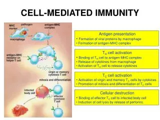
B – CELL ACTIVATION. Ligand. Ligand. SIGNAL. SIGNAL. RECEPTOR MEDIATED CELL ACTIVATION. Cross - linking. C onformational change. CROSS – LINKING OF THE RECEPTOR INITIATES A SIGNALING CASCADE. ligand. kinase activation. phosphorylation. r ecruitment o f adaptors.

E N D
Ligand Ligand SIGNAL SIGNAL RECEPTOR MEDIATED CELL ACTIVATION Cross - linking Conformational change
CROSS – LINKING OF THE RECEPTOR INITIATES A SIGNALING CASCADE ligand kinase activation phosphorylation recruitmentof adaptors Activation of transcription factors Gene transcription SIGNAL
BCR signaling (review)
Ig domain + CHO a b ITAM ITAM Y Y Y Y SIGNALING UNITS OF THE B-CELL RECEPTOR Ig-a/CD79a Ig-b/CD79b ITAM: YxxLx7YxxI ITAM: Immunoreceptor Tyrosine-based Activation Motif
KINETICS OF LYMPHOCYTE ACTIVATION Nyugvó limfocita G0 Resting lymphocyte G0 Co-receptor Adhesion molecule Cytokines SIGNAL2. Effector cellMemory cell Transport Membrane change RNA and protein synthesis proliferation DNA synthesis Lymphoblast PTK activation RNA synthesis Free Ca++ Protein synthesis Protein phosphorylation DNA synthesis Resting lymphocyte G0 0 10sec 1min 5min 1hr 6 hrs 12 hrs 24 hrs ANTIGEN SIGNAL1.
THE CO-STIMULATORY ROLE OF CR2 (CD21) COMPLEMENT RECEPTOR IN B – LYMPHOCYTES C3d ANTIGEN Antigenic determinant CD21 CD19 TAPA=CD81 B-CELL Y Y Enhanced B-cell activation
Mannose Tissue cells Bacterium Antigen B Cell THE NEURAMIC ACID RECEPTOR CD22 INHIBITS ACTIVATION THROUGH THE A B-CELL RECEPTOR Neuraminic (sialic) acid CD22 Inhibited B cell activation
STRUCTURE OF IMMUNOGLOBULINS/ANTIBODIES Heavy chain (H) VH VL CH Light chain (L) CL Antigen Antigen binding Variable domains COMPLEMENT ACTIVATION BINDING TO CELLS DEGRADATION TRANSPORT Constans domains Effector functions
AMINO ACID SEQUENCE OF IMMUNOGLOBULINS Multiple myeloma (MM) Plasma cell tumors – tumor cells reside in the bone marrow Produce immunoglobulins of monoclonal origin,serum concentration 50-100mg/ml Rodney Porter & Gerald Edelman 1959 – 1960 myeloma protein purification L H Reduction 1 2 3 4 5 6 7 8 9 10 11 12 13 14 15 16 17 18 Variable Constant Gel electrophoresis 50 kDa Heavy chain 25 kDa Light chain
GENETIC BACKGROUND OF ANTIBODY DIVERSITY VH VH VL VL S – S S – S Mechanism of the generation of variability? Different rules for encoding the variable and constant regions? Symmetric molecule two identical VH and VL both chromosomes encode for the same sequence?
MOLECULAR GENETICS OF IMMUNOGLOUBLINS How can the bifunctional nature of antibodies be explained genetically? In 1965, Dreyer & Bennett proposed that for a single isotype of antibody there may be: • A single C region gene encoded in the GERMLINE and separate from the V region genes • Multiple choices of V region genes available • A mechanism to rearrange V and C genes in the genome so that they can fuse to form a complete Immunoglobulin gene. This was genetic heresy as it violated the then accepted notion that DNA was identical in every cell of an individual
A single C region gene is encoded in the germline and separated from the multiple V region genes V V V V V V V V V C V V V C A mechanism to rearrange V and C genes in the genome exists so that they can fuse to form a complete Immunoglobulin gene V V Proof of the Dreyer - Bennett hypothesis Find a way to show the existence of multiple V genes and rearrangement to the C gene
V V Germline DNA V V V V V V V C V V V C Rearranged DNA V V Approach • Tools: • A set of cDNA probes to specifically distinguish V regions from C regions • DNA restriction enzymes to fragment DNA • Examples of germline (e.g. placenta) and mature B cell DNA (e.g. a plasmacytoma/myeloma)
DOGMA OF MOLECULAR BIOLOGY CHARACTERISTICS OF IMMUNOGLOBULIN SEQUENCE 1 GEN = 1 PROTEIN THEORIES 1 GEN High rate of somatic mutations in the V-region Gen V C Many GENES (10000 – 100000) Protein V1 C V2 C Vn C
Kb V-probe 6,0 V C-probe 4,0 C 1,5 VC 6.0 Kb 4.0 Kb V V C C 1.5. Kb Experiment of Susumi Tonegawa 1975 Basel DNA-extraction Digestion by restriction enzyme Gel electrophoresis Southernblot B-cell Liver cell B-cell
Gén V and C genes get close to each other in B-cells only V V V V V V V C C B-CELL GÉN SZEGMENSEK SZOMATIKUS ÁTRENDEZŐDÉSE EGY GÉNNÉ Fehérje CONCLUSION There are many variable genesbut only one constant gene GERM LINE
L VL JL CL L VL CL ~ 95aa ~ 100aa ~ 95aa ~ 100aa VL CL L Some of the extra amino acids are provided by one of a small set of J or JOINING regions ~ 208aa Ig gene sequencing complicated the model The structures of germline VL genes were similar for Vk, and Vl, However there was an anomaly between germline and rearranged DNA: Where do the extra 13 amino acids come from?
L VL JL CL Further diversity in the Ig heavy chain DH JH L VH CH The heavy chain was found to have further amino acids (0 – 8) between the JH és CH genes D (DIVERSITY) region Each heavy chain requires 3 recombination events JH to DH, VH to JHDH,and VHJHDH to CH Each light chain requires 2 recombination events VL to JL and VLJL to CL
IMMUNOGLOBULIN CHAINS ARE ENCODED BY MULTIPLE GENE SEGMENTS Gene segments Light chain Heavy chain kappa lambda Variable (V) 132/40 105/30 123/65 Diversity (D) 0 0 27 Joining (J) 5 4 9 ORGANIZATION OF IMMUNOGLOBULIN GENE SEGMENTS Chromosome 2 kappa light chain gene segments Chromosome 22 lambda light chain gene segments Chromosome 14 heavy chain gene segments HOW MANY IMMUNOGLOBULIN GENE SEGMENTS
SOMATIC REARRANGEMENT OF KAPPA (κ) CHAIN GENE SEGMENTS B-cell 2 4 Jκ 80 Vκ Germ line Vκ Jκ Jκ Jκ Jκ Vκ Vκ Vκ Vκ Vκ Vκ Vκ During B-lymphocyte development Jk Jκ Jκ Jκ B-cell 1 Jκ DNA
Leader pA Vκ P Vκ Vκ Vκ Vκ J J J J J J E E Vκ-Jκ Primary RNAtranscript Cκ Cκ Cκ Cκ mRNA AAAA Translation Protein EXPRESSION OF THE KAPPA CHAIN Efficiency of somatic gene rearrangement?
SOMATIC REARRANGMENT OF THE HEAVY CHAIN GENE SEGMENTS During B-cell development VH1 VH1 VH1 D D D D D D D D JH JH VH2 JH JH VH2 VH3 120 VH 12 D 4 JH VH2 VH3 JH JH JH JH
VARIABILITY OF B-CELL ANTIGEN RECEPTORS AND ANTIBODIES VH JH D V-Domains C-Domains VL JL VH-D-JH VL-JL B cells of one individual 1 2 3 4
ORDER OF REARRANGEMENTS OF IMMUNOGLOBULIN GENE SEGMENTS Surrogate light chain D – J recombination V – DJ recombination V – J recombination VDJ –δ transcription δ translation VJ – (or VJ - ) transcription or translation B-sejt Secreted IgM mIgD mIgM
Estimates of combinatorial diversity Taking account of functional V D and J genes: 40 VH x 27 DH x 6JH = 6,480 combinations D can be read in 3 frames: 6,480 x 3 = 19,440 combinations 29 Vk x 5 Jk = 145 combinations 30 Vl x 4 Jl = 120 combinations = 265 different light chains If H and L chains pair randomly as H2L2 i.e. 19,440 x 265 = 5,151,600 possibilities Due only to COMBINATORIAL diversity In practice, some H + L combinations do not occur as they are unstable Certain V and J genes are also used more frequently than others. There are other mechanisms that add diversity at the junctions between genes - JUNCTIONAL diversity GENERATES A POTENTIAL B-CELL REPERTOIRE
THE RESULT OF SOMATIC GENE REARRANGEMENTS • Combination of gene segments results in a huge number of various variable regions of the heavy and light chains expressed by different B-cells • SOMATIC GENE REARRANGEMENT • 2. Successful somatic rearrangement in one chromosome inhibits gene rearrangement in the other chromosome • ALLELIC EXCLUSION • 3. One B-cell produces only one type of heavy and one type of light chain • COMMITMENT TO ONE TYPE OF ANTIGEN BINDING SITE • 4. The B-cell pool consist of B-cells with differently rearranged immunoglobulin genes INDEPENDENT OF ANTIGEN OCCURS DURING B-CELL DEVELOPMENT IN THE BONE MARROW
Y a B a/a Y b B b/b Y a B Y Y Y b a B B b a/b Evidence for allelic exclusion ALLOTYPE- a polymorphism in the Heavy chain C region of Ig Allotypes can be identified by staining B cell surface Ig with antibodies AND Suppression of H chain rearrangement by pre-B cell receptor prevents expression of two specificities of antibody per cell
S. typhi S. typhi Allelic exclusion is needed for efficient clonal selection Antibody All daughter cells must express the same Ig specificity otherwise the efficiency of the response would be compromised Suppression of H chain gene rearrangement helps to prevent the emergence of new daughter specificities during proliferation after clonal selection
Y Y Y Y Y B Self antigen expressed by e.g. brain cells B Y Y Y Y S. aureus S. aureus Y Y Y Y Y Y Y Y Y Y Y Y Anti S. aureus Antibodies Anti S. aureus Antibodies Anti brain Abs Y Y Y Y Y Y Y Y Allelic exclusion prevents unwanted responses One Ag receptor per cell IF there were two Ag receptors per cell Suppression of H chain gene rearrangement ensures only one specificty of Ab expressed per cell. Prevents induction of unwanted responses by pathogens
One specificity of Agreceptor per cell IF there were two specificitiesof Ag receptor per cell Y Y Y S. aureus Y B B Y B B B B Y OR Y Y B B Deletion Anergy Allelic exclusion is needed to prevent holes in the repertoire Anti-brain Ig Anti-brain Ig AND anti-S. Aureus Ig Exclusion of anti-brain B cells i.e. self tolerance anti S.Aureus B cells will be excluded leaving a “hole in the repertoire” BUT
SYNTHESIS OF IMMUNOGLOBULINS Secreted Ig Membrane Ig Golgi ER H and L chains are synthesized on separated ribosomes CHAPERONES Leader sequence Ribosome mRNA