Download
1 / 68
680 likes | 1.34k Views
Case Study. A 26 year old female patient was referred to your clinic from pulmonology after pulmonary function tests indicated a fixed, extrathoracic, airway obstruction.. Case Study. The patient complained of dyspnea on exertion.She denied stridor or voice change.She had been treated for about 1 year by her family doctor for presumed asthma..

E N D
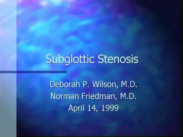
1. Subglottic Stenosis in Wegenger�s Granulomatosis Chad Simon, MD
Susan McCammon, MD
University of Texas Medical Branch
Department of Otolaryngology
Grand Rounds Presentation
May 21, 2008
2. Case Study A 26 year old female patient was referred to your clinic from pulmonology after pulmonary function tests indicated a fixed, extrathoracic, airway obstruction.
3. Case Study The patient complained of dyspnea on exertion.
She denied stridor or voice change.
She had been treated for about 1 year by her family doctor for presumed asthma.
4. Case Study Review of systems was positive for night sweats, achy joints, and nasal congestion.
Her past medical history was positive for recurrent sinusitis and epistaxis.
Family and social history were unremarkable.
She used an albuterol inhaler as needed for shortness of breath.
She had no drug allergies.
5. Case Study Physical exam revealed nasal septal perforation on anterior rhinoscopy.
6. Case Study Physical also revealed bilateral otitis media with effusion and mild conjunctivitis.
7. Case Study Fiberoptic exam revealed a 60% (Cotton-Myer grade II) subglottic stenosis with the appearance of a mature scar.
8. Outline Overview
Pathogenesis
Epidemiology
Pathophysiology
Diagnosis
Treatment
9. Overview In 1931, Heinz Klinger of the University of Berlin first reported two patients who died having prolonged sepsis with inflammation of blood vessels scattered throughout the body.
Five years later, Friederic Wegener in Breslau described a distinct syndrome in three patients.
These patients were found to have necrotizing granulomas involving the upper and lower respiratory tract.
In 1954, seven more patients were described, resulting in the establishment of the definite criteria for the diagnosis of the disease described by Wegener.
10. Overview Wegener joined the Nazi Party in 1932. As a high ranking military doctor he spent some of WWII in a medical office near a Jewish ghetto in Lodz, Poland.
There is speculation that he participated in experiments on concentration camp inmates.
After his Nazi past was discovered in 2000, the chest physician group began a movement to rename Wegener's granulomatosis.
Dr. Friederic Wegener died in July of 1990 at the age of 83.
11. Overview Wegener�s Granulomatosis is a necrotizing granulomatous vasculitis of autoimmune origin
The disease has a predilection for the upper and lower respiratory tracts and the kidneys
In the sinonasal tract, the vasculitis can cause sinusitis, nasal crusting, epistaxis, septal perforation and saddle-nose deformity.
In the lungs, the disease may lead to pneumonitis and hemoptysis.
In the kidneys, a crescentic glomerulonephritis develops, eventually leading to renal failure.
Other manifestations include skin lesions, arthritides, conjunctivitis, and other non-specific systemic inflammatory problems.
13. Pathologic findings. (a) There is swelling and erythema of the nose extending to the upper lip. The external nasal septum appears ulcerated with purulent exudate. (b) The nasal biopsy (performed on the left nares) reveals an atypical dense infiltrate of medium-sized lymphoid cells admixed with a few smaller cells (H&E, original magnification 400). (c) A representative glomerulus shows compression of the tuft by a large cellular crescent admixed with fibrin. In the distorted tuft, there is global mesangial and segmental endocapillary hypercellularity. (H&E, original magnification 400). (d) Immunofluorescence staining shows intense positivity for IgA in a global mesangial distribution (original magnification 400).Pathologic findings. (a) There is swelling and erythema of the nose extending to the upper lip. The external nasal septum appears ulcerated with purulent exudate. (b) The nasal biopsy (performed on the left nares) reveals an atypical dense infiltrate of medium-sized lymphoid cells admixed with a few smaller cells (H&E, original magnification 400). (c) A representative glomerulus shows compression of the tuft by a large cellular crescent admixed with fibrin. In the distorted tuft, there is global mesangial and segmental endocapillary hypercellularity. (H&E, original magnification 400). (d) Immunofluorescence staining shows intense positivity for IgA in a global mesangial distribution (original magnification 400).
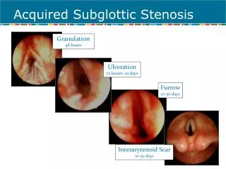
14. Overview Subglottic stenosis is a potentially life-threatening manifestation of Wegener�s granulomatosis.
This narrowing of the upper airway at the level of the cricoid cartilage and/or upper tracheal rings presents a management dilemma.
16. Epidemiology Reported to occur in 16-23% of patients with the disease. (Langford)
Has been reported to occur more often in females. (Shokkenbroek, Gluth)
Median age at diagnosis is 26. (Langford)
Is more likely to occur in patients diagnosed with Wegener�s before age 20. (Langford)
17. Epidemiology Patients with WG and SGS tend to have more sinus involvement and saddlenose deformity than other WG patients.
On the contrary, SGS patients tend to have less lung and kidney involvement. (Langford)
18. Table 1
19. Pathophysiology
The pathogenesis is unclear.
Subglottic stenosis can progress in the absence of systemic disease.
Few patients have WG exacerbations around the time of diagnosis with SGS.
In series where patients were followed, need for repeat procedures was not related to repeated systemic disease flares.
It is postulated that during flares of systemic disease, subclinical subglottic involvement occurs, which subsequently heals with circumferential scarring. (Shokkenbroek)
20. Pathophysiology It is postulated that the subglottis is vulnerable because it is a watershed area of the microcirculation.
This watershed area is the junction of 2 separate embryological growth centers. (Eliachar)
Exposure of respiratory epithelium to gastric contents during LPR episodes is also believed to play a role. (Gluth)
Initial granulomatous inflammation is followed by circumferential scarring and airway narrowing.
22. Diagnosis Dyspnea on exertion is the most common presenting symptom. (79-82%)(Gluth, Langford)
Other symptoms include voice change, stridor, or cough.
Patients may have a known diagnosis of WG (50%), have other symptoms of systemic disease, or present as a new patient with airway complaints.
23. Diagnosis If the diagnosis of WG is not established, an anti-cytoplasmic antibody assay should be performed.
It has been suggested that ANCA titers be performed on nearly all patients with SGS. (Gluth)
24. Diagnosis A positive C-ANCA assay is reported to be 91% sensitive and 99% specific for active Wegener�s granulomatosis.
However, a series of patients with SGS and presumed WG showed that only 57% initially showed a positive ANCA assay.
85% of the cohort eventually became positive at a later date. (Alaani)
This emphasizes the fact that SGS can be present in the absence of active disease.
It also suggests that ANCA assay be repeated serially if there is diagnostic uncertainty.
25. Diagnosis The presence of ANCA in serum can be detected by indirect immunofluorescence or by radioimmunoassay.
Indirect immunofluorescence reveals a pattern of staining that can be more specific for a particular disease.
The �cytoplasmic� pattern (c-ANCA) is associated with ANCA reacting with a 29 kD protein from the azurophilic granules of the neutrophil.
The �perinuclear� (p-ANACA) pattern is associated with ANCA reacting with myeloperoxidase.
Radioimmunoassay of ANCA titers can be used as an initial screening test and to monitor therapy and detect flare-ups of systemic disease.
26. (A) C-ANCA with cytoplasmic staining, central accentuation and PR3 specificity.
27. (C) P-ANCA with perinuclear fluorescence and nuclear extension, where the antigen was MPO.
28. Diagnosis The airway should be fiberoptically examined in the clinic setting.
Topical laryngeal anesthesia may be necessary to visualize the sublottic lesion.
In the majority of cases (75%), the stenotic segment has the appearance of a mature scar and lacks acute inflammatory changes. (Gluth)
CT scans will help to delineate the extent of the lesion but should not be used for primary diagnosis.
30. Diagnosis
31. Diagnosis The stenosis can be graded using the Cotton-Myer classification scheme for grading circumferential subglottic stenosis.
Grade I - Obstruction of 0-50% of the lumen obstruction
Grade II - Obstruction of 51-70% of the lumen
Grade III - Obstruction of 71-99% of the lumen
Grade IV - Obstruction of 100% of the lumen (ie, no detectable lumen)
32. Diagnosis Histological proof is obtained by finding vasculitis, necrotizing granulomata and giant cells in biopsy material.
In general, biopsies of the subglottic lesion are not sensitive for the detection of WG.
Only 5-15% of subglottic biopsies performed on patients with positive ANCA titers and SGS return changes consistent with WG. (Gluth, Langford)
In contrast, nasal biopsies on a similar cohort of patients yielded 82% sensitivity for WG. (Gluth)
33. Paranasal sinus tissue reveals a florid mixed inflammatory cell infiltrate in a granulation tissue-type background. Inflammatory cells include multinucleated giant cells and neutrophil microabscesses (A, H&E, �200).
34. Diagnosis Pulmonary function tests are abnormal in 60% of patients with subglottic stenosis. (Langford)
Flattening occurs in the flow-volume loop in both the inspiratory and expiratory portions, giving it a box-like appearance.
However, PFTs may not detect less severe stenoses and should never be used for primary diagnosis.
An abnormal flow-volume loop is correlated with need for surgical intervention. (Langford)
35. Figure 2
36. Treatment Treatment of subglottic stenosis should be considered based on the presence of symptoms combined with objective evidence of tracheal narrowing.
Other parameters, such as active systemic disease or a change in ANCA titers should never be used as consideration for procedures.
37. Treatment Immunosuppression
Tracheostomy
Endoscopic dilation
Intralesional steroids
Laser procedures
Cold knife lysis
Open surgical procedures
Stents
38. Immunosuppression Drugs such as corticosteroids, cyclophosphamide, and azothioprine has revolutionized the treatment of WG, with a dramatic reduction in mortality.
However, subglottic lesions are generally unresponsive to systemic agents.
49% of SGS cases are diagnosed while a patient is on active therapy. (Langford)
Success rates of medical therapy in relieving the obstruction vary from 22%-26%. (Gluth, Langford)
39. Tracheostomy Life-threatening airway obstruction may require tracheostomy as a temporizing or permanent measure.
Tracheostomy is necessary in 8 - 60% of cases. (Langford, Schokkenbroek, Gluth, Alaani)
Often, success of other therapies is measured by decannulation of the tracheostoma.
40. Endoscopic Dilation Endoscopic procedures have been variably successful at managing this airway lesion.
Dilation tracheoscopy can be performed using a Groningen optical dilatation tracheoscope (Karl Storz 1033R)(Fig 1) fitted with a 30 cm Hopkins telescope (shown below).
41. Endoscopic Dilation Under general mask anesthesia, the scope is introduced.
The beveled, fenestrated tip is designed to ensure ventilation as the scope is advanced through the stenosis.
The conical design allow the scope to be advanced up to its wider portion.
The scope is left in place for 5 � 10 minutes.
43. Endoscopic Dilation Dilation tracheoscopy has been shown to be effective the majority of the time for Cotton-Meyer grade I-II in the short term.
Repeat procedures are commonly necessary.
No complications have been reported with this procedure.
The small number of reported cases (9) should be considered. (Shoekkenbroek)
44. Endoscopic Dilation
45. Endoscopic Dilation with Steroid Injection Traditional dilation techniques, combined with intralesional steroid injection have been reported.
Under suspension laryngoscopy and spontaneous ventilation, graduated dilators are used to dilate the trachea.
Next, methylprednisolone injections are performed in a a 4-quadrant, submucosal pattern.
Perioperative, systemic steroids are also used.
47. Endoscopic Dilation with Steroid Injection 20 patients received this treatment and were followed for a median of 35 months.
The median number of treatments required was 3.
Six patients required only 1 procedure.
One patient required 22 procedures.
All patients who began therapy with a tracheostomy were eventually decannulated.
No patients required a new tracheostomy.
48. Endoscopic Dilation with Steroid Injection In another series, 21 patients with WG and significant SGS were studied.
These patients were treated with intralesional steroid injection combined with mechanical dilation and cold-knife lysis of the lesion.
Under suspension laryngoscopy and jet ventilation, methylprenisolone was injected submucosally in 4 quadrants.
49. Endoscopic Dilation with Steroid Injection Lysis of the stenotic ring was then performed by making radial incisions with a laryngeal microsickle knife.
The stenosis was then serially dilated with Maloney bougies or a Foley catheter.
Topical mitomycin C was variably used. (Hoffman)
50. Endoscopic Dilation with Steroid Injection Patients with prior procedures (mostly laser) averaged more procedures to obtain adequate airway patency and with a shorter interval between procedures.
The authors comment that, subjectively, lesions seen in patients who had undergone prior laser procedures were severe, extensive, and thickly fibrotic.
No new tracheotomies were necessary in either group. (Hoffman)
52. Endoscopic Dilation with Steroid Injection The most difficult lesions were found in the 6 patients with prior tracheotomies, presenting with multilevel stenoses, cicatrix formation, vocal cord fixation and arytenoid damage.
In these patients, widening of the subglottic region alone was not sufficient.
4 of the 6 patients achieved decannulation by means of various laryngotracheoplastic techniques which were not specified.
53. Endoscopic Dilation with Steroid Injection The authors comment that laser procedures cause extensive scarring and thus patients are more difficult to manage afterward.
However, these patients may represent a cohort with more severe disease, who would be more difficult to manage anyway.
There is no way to differentiate whether the scarring is from prior laser procedure, or from more extensive inflammation in the airway .
54. Endoscopic Dilation with Laser Treatment Although abhorred by other authors, laser treatment of SGS has been performed and reported on.
For lesions close to the vocal cords, in 2 patients, CO2 laser was used with microsuspension laryngoscopy to resect the stenosis.
For lesions far from the cords (>1 cm), in 3 patients, Nd:YAG laser was used with through a fiberoptic bronchoscope, under local anesthesia. (Shvero)
55. Endoscopic Dilation with Laser Treatment 4 out of these 5 patients are reported to have favorable outcomes or decannulation during their follow up period of 6-60 months.
However, all patients required multiple treatments (2-18)
In addition, one patient required placement of a subglottic stent. (Shvero)
57. Laryngotracheal Reconstruction Open surgical procedures have been performed with success.
Most commonly performed is the laryngotracheal reconstruction with costal cartilage graft (LTR).
It is recommended to defer LTR until a patient is in a quiescent phase of the systemic disease.
This should be monitored by serum ANCA titers, CRP, and ESR.
It is also recommended that surgery be deferred until a patient is not actively taking steroids or other immunosuppressant medications. (Gluth)
58. Laryngotracheal Reconstruction A small series of Wegener�s patients who received laryngotracheal reconstruction for SGS shows excellent results.
6 of 6 patients were decannulated from their tracheostomy after the LTR.
There were no cases of graft failure even in 3 of the patients in whom reactivation of systemic WG occurred.
1 complication of graft displacement occurred. (Gluth)
59. Stents The use of airway stents to maintain airway patency in SGS and WG is controversial.
Some authors have advocated deployment of a Nitinol (nickel-titanium alloy) expandable stent into the subglottic airway when all other therapies fail.
Similar stents have been used with success in the distal tracheobronchial tree.
60. Stents
61. Stents A case report exists in the literature.
A patient was followed for 48 months after deployment of a nitinol stent.
She had a patent airway, no complications, and no ingrowth of granulation tissue. (Watters)
62. Stents Other authors argue that the long-term safety and efficacy of these stents has not been well established.
They argue that these devices are associated with stent fracture, migration, increased granulation tissue, and even death.
Stents in the subglottis also undergo movement with swallowing and saliva exposure.
These factors preclude early mucosalization of the stent, as occurs in more distal portions of the airway.
The foreign body reaction, it is argued, causes more granulation tissue ingrowth. (Mair- anecdotal)
63. Stents The argument against stents is further supported by the fact that an emergency trachotomy in the setting of a subglottic metal stent is very difficult to perform.
65. Treatment Plan If airway is tenuous, perform emergent tracheotomy.
If patient is stable, perform PFTs, CT scan, fiberoptic endoscopy.
1st line therapy should be endoscopic dilation.
With subsequent dilations, cold-knife lysis, and intralesional steroids should be added.
Patient should be followed every 3 months with repeat fiberoptic exams and PFTs.
66. Treatment Plan Serial ANCA titers are not necessary from an ENT standpoint, since they correlate poorly with SGS progression.
Endoscopic procedures should be repeated as necessary.
If endoscopic procedures must be performed at an increasingly more frequent interval, consideration should be given to open laryngotracheal reconstruction.
Subglottic stents should be avoided if possible.
67. Outline Overview
Pathogenesis
Epidemiology
Pathophysiology
Diagnosis
Treatment
68. Bibliography Alaani et al. Wegener's granulomatosis and subglottic stenosis: management of the airway. J Laryngol Otol. 2004 Oct;118(10):786-90.
Eliachar et al. New approaches to the management of subglottic stenosis in Wegener's granulomatosis. Cleve Clin J Med. 2002;69 Suppl 2:SII149-51.
Gluth et al. Subglottic stenosis associated with Wegener's granulomatosis. Laryngoscope. 2003 Aug;113(8):1304-7.
Hoffman et al. Treatment of subglottic stenosis, due to Wegener's granulomatosis, with intralesional corticosteroids and dilation. J Rheumatol. 2003 May;30(5):1017-21.
Langford et al. Clinical features and therapeutic management of subglottic stenosis in patients with Wegener's granulomatosis. Arthritis Rheum. 1996 Oct;39(10):1754-60.
Shokkenbroek et al. Dilatation tracheoscopy for laryngeal and tracheal stenosis in patients with Wegener's granulomatosis. Eur Arch Otorhinolaryngol. 2008 May;265(5):549-55. Epub 2007 Nov 14.
Shvero et al. Endoscopic laser surgery for subglottic stenosis in Wegener's granulomatosis. Yonsei Med J. 2007 Oct 31;48(5):748-53.
Watters et al. Subglottic stenosis in Wegener's granulomatosis and the nitinol stent. Laryngoscope. 2003 Dec;113(12):2222-4.