Download

1 / 90
920 likes | 1.16k Views
Overview. Pediatric subglottic stenosisPatient presentation and work-upMedical managementSurgical intervention. Stridor. A harsh, high pitched musical sound that results from turbulent airflow through the upper airwayEtiology may range from mild illness to severe, life-threatening situation. Str

E N D
1. Surgical Management of Pediatric Subglottic Stenosis Michael Briscoe Jr., MD
Seckin Ulualp, MD
UTMB Department of Otolaryngology Grand Rounds
June 27, 2007
2. Overview Pediatric subglottic stenosis
Patient presentation and work-up
Medical management
Surgical intervention
3. Stridor A harsh, high pitched musical sound that results from turbulent airflow through the upper airway
Etiology may range from mild illness to severe, life-threatening situation
4. Stridor Etiology Congenital
Inflammation
Trauma
Foreign bodies
5. Stridor Presentation Variable age of onset
Patient typically presents with sudden onset of symptoms
Acquired stridor (inflammation, trauma, foreign bodies) is more likely than congenital stridor to require airway intervention
6. Congenital Stridor Eighty-five percent of children under 2.5 years presenting with stridor have a congenital etiology
Often not present at birth
Typically presents prior to four months of age
7. Assessing Stridor Determination of respiratory phase in which sound is noted
Inspiratory
Biphasic
Expiratory
8. Inspiratory Stridor Result of supraglottic obstruction
High-pitched
9. Biphasic Stridor Result of extrathoracic tracheal obstruction including
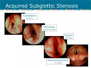
Glottis
Subglottis
Intermediate pitch
10. Expiratory Stridor Result of intrathoracic tracheal obstruction
Associated with prolonged expiratory wheezing
11. Congenital subglottic stenosis Third most common cause of stridor in the neonate.
Involves narrowing of the subglottic lumen in the absence of trauma.
Full term neonate with lumen of less than 4 mm, or 3 mm in premature infant.
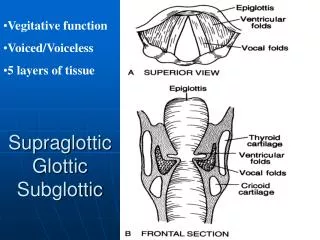
12. Subglottic airway The subglottic airway is the narrowest area of the newborn�s airway since it is a complete, nonpliable, nonexpandable ring.
13. Subglottic stenosis Caused by failure of the trachea to recannalize during embryogenesis.
Approximately 5% of children will require smaller size ET tube than expected for age.
Children with Down syndrome have high incidence of congenital airway narrowing.
14. Subtypes Membranous � usually circumferential, soft and dilatable.
Submucosal fibrosis
Submucosal gland hyperplasia
Granulation tissue
Cartilaginous � has a more variable appearance.
Normal shaped cricoid with decreased lumen
Abnormally shaped cricoid with lateral shelves
Elliptical shape
15. Patient Presentation If severe, will have respiratory distress at birth.
In milder cases, may present in first few weeks of life.
Most become symptomatic by three months of age due to increased activity and ventilatory requirements.
16. Patient Presentation cont. Symptoms of upper airway obstruction predominate.
Inspiratory stridor progresses to biphasic as obstruction worsens.
Exacerbated by URI, and these children tend to have recurrent croup
17. Office evaluation Onset, duration, severity, feeding abnormalities, failure to thrive, recurrent URI/croup
Positional effects on stridor
Voice quality
Symptoms of reflux
Complete head and neck exam
Flexible laryngoscopy
18. Diagnosis Differential
Congenital
Laryngeomalacia
Tracheomalcia
VC paralysis
Cysts
Clefts
Vascular compression
Mass
19. Diagnosis Differential
Infection/Inflammation
Croup
GER
Tracheitis
Neoplastic
Subglottic hemangioma
Recurrent respiratory papillomas
Foreign body
20. Flexible Laryngoscopy Best performed with
Unanesthetized child
Upright position
1.9mm laryngoscope
Scope should be passed through both nasal passages
Evaluate vocal cord mobility
21. Definitive diagnosis Rigid endoscopy
Imaging studies usually not necessary.
Neck films
Flouroscopy
CT/MRI
22. Rigid endoscopy Evaluate the subglottis and glottis for fixation, scarring, granulation, edema, paralysis or paresis, and other abnormalities. Evaluate the distance and caliber of the stenosis. Apply the Myers and Cotton staging system only to circumferential SGS. Glottic stenosis and SGS often coexist and must be considered when reconstruction is planned
23. Grading Systems for SGS
Cotton-Myer (1994)
McCaffrey (1992)
24. Grading Systems for SGS Cotton-Myer
Based on relative reduction of subglottic cross-sectional area
Good for mature, firm, circumferential lesions
Does not take into account extension to other subsites or length of stenosis
25. Cotton-Myer
26. Cotton-Myer ET tubes of various sizes are placed sequentially.
Leak test performed
When 10-25 cm H2O leak pressure achieved, this is patient�s tube size.
Compare to normal values for age.
27. McCaffrey System The McCaffrey system classifies laryngotracheal stenosis based on the subsites involved and the length of the stenosis. Four stages are described: stage I lesions are confined to the subglottis or trachea and are less than 1cm long, stage II lesions are isolated to the subglottis and are greater then 1 cm long, stage III are subglottic/tracheal lesions not involving the glottis, and stage IV lesions involve the glottis
28. Grading Systems for SGS McCaffrey
29. Management of SGS Medical
Observation
Tracheostomy
Airway expansion procedure
30. Management of SGS Medical
Diagnosis and treatment of GER
Pediatric � consultation with primary physician and specialists (pulmonary, GI, cardiology etc.)
Adult
Assess general medical status
Consultation with PCP and specialists
Optimize cardiac and pulmonary function
Control diabetes
Discontinue steroid use if possible before LTR
31. Management of SGS Observation
Reasonable in mild cases, esp. congenital SGS (Cotton-Myer grade I and mild grade II)
If no retractions, feeding difficulties, or episodes of croup requiring hospitalization
Follow growth curves
Repeat endoscopy q 3-6 mo
32. Management of SGS Tracheostomy
Often the initial step in treatment of pediatric acquired SGS
May be avoided in patients with congenital SGS
Allows time for the infant to mature
Lungs � BPD
Wt. � 10 kg (Cotton)
2%-5% mortality in children
Accidental decannulation and plugging
33. Grade I stenosis Usually will grow out of stenosis.
Treatment is medical
May have recurrent croup
If surgery needed, may try endoscopic, dilation, or laser (CO2 or KTP)
34. Grade II Surgery is needed secondary to respiratory distress.
May try endoscopic, dilation, or laser.
May require open procedure
35. High Grade Refers to grade III or IV lesions
Laryngotracheal reconstruction
Anterior
Anterior and posterior
Anterior, posterior, and lateral
Partial cricotracheal resection
36. Preoperative planning Treat GER/EER before attempting reconstruction.
Assess full extent of stenosis.
Order CT scan with 3D reformats if total length of stenosis remains undetermined after rigid bronchoscopy.
Treat any respiratory infections with antibiotics, and steroids
37. Evaluation for reflux Signs of extraesophageal reflux are noted, and include post-cricoid edema, ventricular effacement, and follicular bronchitis.
BAL for lipid-laden macrophages
39. Intervention Goal of intervention is
to have an adequate airway to allow for normal activity without the need for tracheostomy
Single stage procedure, or two stage procedure with minimal postoperative morbidity, and minimal hospital stay. (Cable et al)
40. Cotton�s Stages of Reconstruction Stage 1 � complete evaluation of the airway
Stage 2 � expansion of the subglottic lumen
with preservation of function
Stage 3 � stabilization of the expanded
lumen framework (grafts and/or stents)
Stage 4 � healing
Stage 5 - decannulation
41. Surgery for SGS I. Endoscopic
Dilation
Laser
II. Open procedure
Expansion procedure (with trach and stent or SS-LTR)
Laryngotracheoplasty
Laryngotracheal reconstruction
42. Management of SGS How do you decide which procedure to perform
Status of the patient
Any contraindications
Absolute
Tracheotomy dependent (aspiration, severe BPD)
Severe GER refractive to surgical and medical therapy
Relative
Diabetes
Steroid use
Cardiac, renal or pulmonary disease
43. Management of SGS Endoscopic
Dilation
Practiced frequently before advent of LTR
Requires multiple repeat procedures
Low success rate but an option for patients who cannot undergo LTR
44. Management of SGS Endoscopic
Laser
66-80% success rate for Cotton-Myer grade I and II stenoses
Factors associated with failure
Previous attempts
Circumferential scarring
Loss of cartilage support
Exposure of cartilage
Arytenoid fixation
Combined laryngotracheal stenosis with vertical length >1cm
45. Laser excision of subglottic web
46. Laser excision of subglottic web
47. Management of SGS
Grade III and IV stenoses require and open procedure
48. Anterior cricoid split Patient weight > 1500 grams
Failure to extubate in identified SGS
Oxygen requirement < 30%
No active respiratory infection
Good pulmonary and cardiac function.
49. ACS
50. ACS Remain intubated 7-10 days
Ab and antireflux meds while intubated.
Complications include reintubation, pneumothorax, pneumomediastinum, subcutaneous emphysema, wound infection, and persistent SGS.
Success rate of 58-100%
51. Single Stage LTR Surgical correction with short period of stenting.
Two stage procedure still necessary for patients with poor pulmonary reserve, or multilevel stenosis.
Grade II and selected grade III SGS.
52. SSLTR Same approach as for ACS
Remove ET tube when air leak at 20 cm H2O.
53. SSLTR Gustafson et al. Retrospective chart review at tertiary care hospital.
200 pediatric patients, 96% decannulation rate.
29% required reintubation, 15% needed trach
4% remained trach dependent
Anterior/posterior vs. anterior or posterior, higher rates of reintubation
70% Grade I/II
EBM C
54. Gustafson et al Age greater than four, less complications after extubation and less need for sedation. (48 hours)
Increased duration of stenting did not affect outcome. Follow leak pressure. 20 cm H2O
Moderate to severe tracheomalacia may be contraindication
55. Complications Younis et al. Retrospective chart review. 46 patients underwent A/P SSLTR. 35 Grade III/VI.
83% decannulation rate
EBM C
56. Posterior SSLTR
57. LTR with stenting Anterior � adequate for isolated anterior subglottic stenosis
Anterior/posterior � for circumferential or posterior SGS
Anterior/posterior/lateral � for complete SGS
58. LTR Introduced in 1972 by Fearon and Cotton.
Widely used
Tracheostomy and stent in place for several months
59. LTR Same approach as ACS.
May perform posterior split if needed. Must be aware of esophageal mucosa to avoid inadvertent injury.
Stenting/tracheostomy short term (4-6 weeks) or long term (>2 months)
60. Duration of stenting Duration of stenting dependent on:
Amount of rigidity in the area of stenosis
Distortion of anatomy
Propensity for keloid formation/hypertrophic scar
Stability of grafts
Scar contracture
61. Complications Dysphagia
Aspiration
Granulation tissue
Dislodgement of stent
62. Granulation tissue
63. Factors leading to failure Choi et al, retrospective chart review at tertiary care children�s hospital.
17 patients requiring 42 LTRs
2 perioperative deaths, 15 successfully decannulated.
27 failed procedures
24 of 27 failed procedures, at least one cause could be found for failure.
EBM-C
64. Factors leading to failure Preoperative
Inadequate assessment of post. SGS
Intraoperative
Stent
Duration, length, type
graft
Postoperative
Keloid formation, GERD, suprastomal/infrastomal collapse, poor follow-up, slipped or broken stent
65. Stents Aboulker, Montgomery T-tube, silastic swiss roll (portex and finger cot - no longer used). All have there own limitations, complications.
Aboulker is rigid, providing stenting and less collapsibility.
Swiss roll causes granulation tissue, gentle pressure.
used less often
Montgomery stent for older children with adequate distance between glottis and stenosis.
Associated plugging, with airway obstruction
Used less often
66. Aboulker and Montgomery stents
67. Aboulker � most frequently used stent
68. Montgomery T tube Lumen with small caliber, easily occluded
Used less frequently than Aboulker stent.
69. Cartilage Cartilage is better material because they have a lower rate of resorption, are easy to carve, and are viable without a vascular pedicle. They also retain bulk even without functional use.
Rib and auricular most commonly used.
Can not use grafts for lateral splits
70. Graft material Auricular cartilage
Thyroid cartilage
Hyoid bone
Rib cartilage
Irradiated cartilage
71. LTR with stenting Procedures requiring long term stenting falling out of favor.
SSLTR or two-stage LTR preferred
CTR another option for high grade stenosis
72. Cricotrachael resection First reported in 1970 and popularized in the 90s.
More technically challenging than split procedures.
Can be used as salvage for failures
73. Success rates White et al, retrospective chart review of 100 consecutive patients at tertiary care center.
96 total patients, 89 with Grade III/IV stenosis
94% decannulation rate
Vocal cord dysfunction was only significant risk factor for failure to decannulate after 1 surgery.
MRSA and pseudomonal infections may play a role in failure, but cohort too small.
EBM-C
74. CTR Best candidates are those with severe SGS, without associated glottic pathology and with at least 4mm in healthy airway below the vocal folds and above the stenosis.
75. Exposure for CTR
76. CTR � Line of resection in relation to recurrent laryngeal nerve
77. Elevation of perichondrium from anterior cricoid arch to avoid recurrent laryngeal nerve injury
78. CTR � anterior cricoid arch excised
79. CTR � removal of soft tissue of posterior cricoid plate
80. CTR � optional partial laryngofissure for increased luminal diameter
81. CTR � dissection of party wall
82. Completed CTR
83. CTR � completed reconstruction with stay sutures
84. CTR � posterior anastamosis
85. CTR
86. CTR complications Anastamosis failure
Granulation tissue
RLN injury
Arytenoid prolapse
Restenosis
Wound infection
Need for further procedures
Re-intubation
87. Postoperative Care Intensive care unit
Intensivist familiar with these cases
Patients with trach and stent
Abx
Antireflux
Trach care teaching
Often discharged in several days
Repeat endoscopy q 3-4 weeks for stent evaluation
Stent duration
Depends on purpose
Hold graft in place � as little as one weeks
Counteract scar formation � months to a year
88. Postoperative Care ACS or SS-LTP
More intense care
Intubated 7-14 days with ETT as stent
Broad spectrum abx
Antireflux
Chest physiotherapy and log rolling
May need paralysis
Extubate when audible air leak at 20 cm H20
Decadron 1mg/kg 12hrs prior to extubation and 5 days postextubation
89. Conclusions Fiberoptic laryngoscopy and direct laryngoscopy/bronchoscopy essential for diagnosis.
Choice of procedure dependent on grade of stenosis, ability of surgeon, and diligent post-operative care.
High decannulation rates, but may require multiple procedures.
90. Bibliography Bath AP, Panarese A, Thevasagayam M, Bull PD. Paediatric subglottic stenosis. Clinical Otolaryngol 1999; 24(2):117-121
Bosely et al. �Pediatric partial CTR: a new technique for posterior anastomisis.� Oto Head and Neck Surgery 2006. 135; 318-322
Cable et al. Pediatric airway reconstruction: Principles in decision-making and outcomes at The University of Iowa Hospitals and clinics. Ann Otolo Rhinol Laryngol 113:2004; 289-293
Choi et al. �Pitfalls in LTR� Arch Otolaryng Head Neck Surgery. 1999 125, 650-53.
Cotton RT, O'Connor DM. Paediatric laryngotracheal reconstruction: 20 years experience. Acta Otorhinolaryngol Belg 1995; 49:367-372
Fayou et al. �Thyroid alar cartilage in paediatric LTR.� International Journal of Pediatric Otorhinolaryngology 2006; 70, 717-724.
Gustafson et al. �SSLTR in children: a review of 200 cases. Otol Head and Neck; Vol.123;4
Halstead LA. Role of Gastroesophageal Reflux in Pediatric Upper Airway Disorders. Otolaryngol Head Neck Surg 1999;120:208
Koltai et al. �Anterior-posterior cartilage graft dimensions in successful LTR� Arch Otolaryng Head Neck Syrgery. 2006;132 631-34
McCaffrey TV. Classification of laryngotracheal stenosis. Laryngoscope 1992:102:335-340
Monnier PM et al. Cricotracheal resection for adult and pediatric subglottic stenoses: similarities and differences. Operative Techniques in Otolaryngol-Head Neck Surg 1999; 10(4): 311-315
Myer CM, O�Connor DM, Cotton RT. Proposed Grading System for Subglottic Stenosis Based on Endotracheal Tube Sizes. Ann Otol Rhinol Laryngol 1994; 103:319
Nitin et al, �The workup of stridor: virtual bronchoscopy� Respiratory Care March 2007. Vol 52 No 3.
White et al. �Pediatric cricotracheal resection.� Arch Otolaryng Head Neck; Oct. 2005 Vol. 131
Younis et al. �Post. Cartilage graft in SSLTR.� Oto Head and Neck Surgery. Vol 129;3 168-175
Zalzal GH. Use of stents in laryngotracheal reconstruction in children: Indications, technical considerations, and complications. Laryngoscope 1988; 98:849-854